Copyright
©2014 Baishideng Publishing Group Inc.
World J Gastroenterol. Jun 14, 2014; 20(22): 7005-7010
Published online Jun 14, 2014. doi: 10.3748/wjg.v20.i22.7005
Published online Jun 14, 2014. doi: 10.3748/wjg.v20.i22.7005
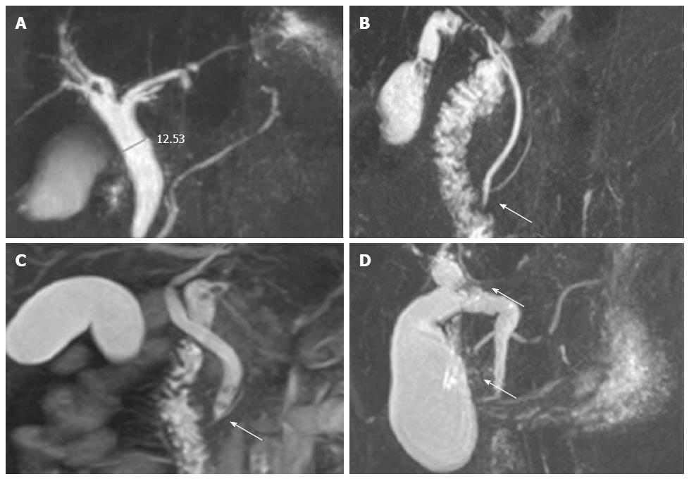
Figure 1 Types of bile duct and pancreatic duct.
A: Normal type of pancreaticobiliary duct union in a 45 years old woman. The common duct can hardly be seen and the common bile duct widens (diameter, 12.53 mm); B: Pancreatic duct joins to common bile duct in a 39 years old man (arrow, P-B type); C: Common bile duct joins to pancreatic duct in a 55 years old woman (arrow, B-P type); D: Common bile duct and pancreatic duct don’t join while opening into duodenal wall separately in a 44 years old woman (arrows).
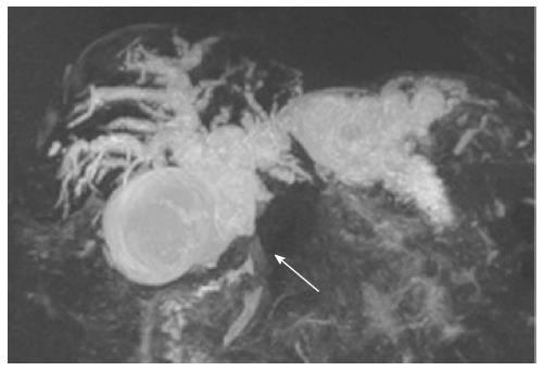
Figure 2 B-P type of pancreaticobiliary maljunction in a 53 year old man with gallbladder carcinoma.
Gallbladder volume enlarges, and the middle part of the common bile duct isn’t displayed (arrow). The length of the common duct is larger than 1 cm (10.07 mm).
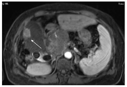
Figure 3 Lateral wall of the gallbladder shows a mass in a 69 year old man with gallbladder carcinoma, with unclear boundary and heterogeneous enhancement growing into capsular space.
An irregular mass (arrow) in the hepatic hila above the pancreatic head shows heterogeneous enhancement after enhanced scan.
- Citation: Wang CL, Ding HY, Dai Y, Xie TT, Li YB, Cheng L, Wang B, Tang RH, Nie WX. Magnetic resonance cholangiopancreatography study of pancreaticobiliary maljunction and pancreaticobiliary diseases. World J Gastroenterol 2014; 20(22): 7005-7010
- URL: https://www.wjgnet.com/1007-9327/full/v20/i22/7005.htm
- DOI: https://dx.doi.org/10.3748/wjg.v20.i22.7005


