Copyright
©2014 Baishideng Publishing Group Inc.
World J Gastroenterol. Jun 7, 2014; 20(21): 6698-6700
Published online Jun 7, 2014. doi: 10.3748/wjg.v20.i21.6698
Published online Jun 7, 2014. doi: 10.3748/wjg.v20.i21.6698
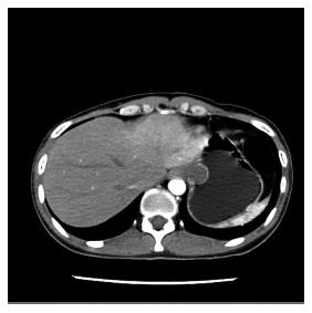
Figure 1 Computed tomography revealed a tumor located in the gastric fundus adjacent to gastric cardiac region.
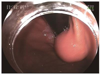
Figure 2 Gastroscopy showed the gastric fundus tumor.
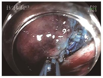
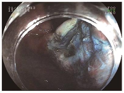
Figure 3 Endoscopic submucosal dissection of the basal area of the tumor.
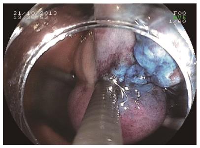
Figure 4 Blunt dissection using the spring rolling pattern.
Figure 5 Base of the tumor after dissection, no perforation can be seen.
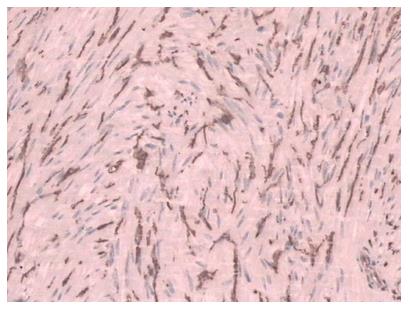
Figure 6 Immunohistochemistry confirmed the diagnosis of gastrointestinal stromal tumor.
- Citation: Wen ZQ, Wu GY, Yu SP, Lin XD, Li SH, Huang XG, Zhang F, Zeng XY, Huang HY, Li AM. Application of blunt dissection in ESD of a gastric submucosal tumor. World J Gastroenterol 2014; 20(21): 6698-6700
- URL: https://www.wjgnet.com/1007-9327/full/v20/i21/6698.htm
- DOI: https://dx.doi.org/10.3748/wjg.v20.i21.6698


