Copyright
©2014 Baishideng Publishing Group Inc.
World J Gastroenterol. Jun 7, 2014; 20(21): 6586-6593
Published online Jun 7, 2014. doi: 10.3748/wjg.v20.i21.6586
Published online Jun 7, 2014. doi: 10.3748/wjg.v20.i21.6586
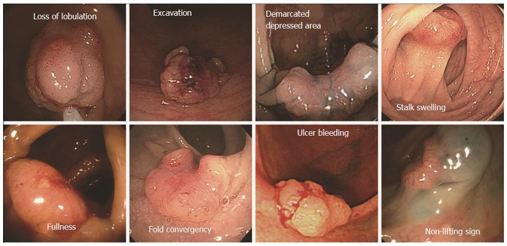
Figure 1 Seven morphological features and non-lifting sign for submucosal cancer.
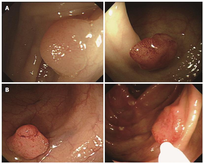
Figure 2 Pit-pattern classification; (A) non-invasive pattern, and (B) invasive pattern.
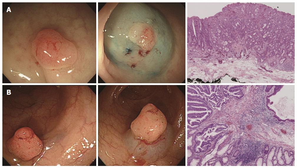
Figure 3 An 8-mm sessile polyp with negative non-lifting signs was treated by endoscopic mucosal resection (A); histology showed that cancer cells invaded the submucosa up to 200 μm (B); a 12-mm sized sessile polyp with negative non-lifting signs was treated by endoscopic mucosal resection.
Histology showed that cancer cells invaded the submucosa to 2000 μm. The patient received additional surgery.
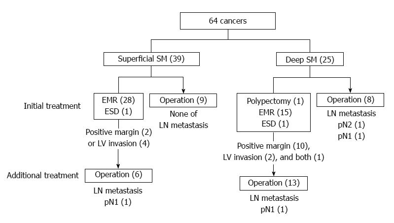
Figure 4 Diagram for the treatment of 64 early colorectal cancers.
SM: Submucosa; EMR: Endoscopic mucosal resection; ESD: Endoscopic submucosal dissection.
- Citation: Park W, Kim B, Park SJ, Cheon JH, Kim TI, Kim WH, Hong SP. Conventional endoscopic features are not sufficient to differentiate small, early colorectal cancer. World J Gastroenterol 2014; 20(21): 6586-6593
- URL: https://www.wjgnet.com/1007-9327/full/v20/i21/6586.htm
- DOI: https://dx.doi.org/10.3748/wjg.v20.i21.6586


