Copyright
©2014 Baishideng Publishing Group Inc.
World J Gastroenterol. May 21, 2014; 20(19): 5859-5866
Published online May 21, 2014. doi: 10.3748/wjg.v20.i19.5859
Published online May 21, 2014. doi: 10.3748/wjg.v20.i19.5859
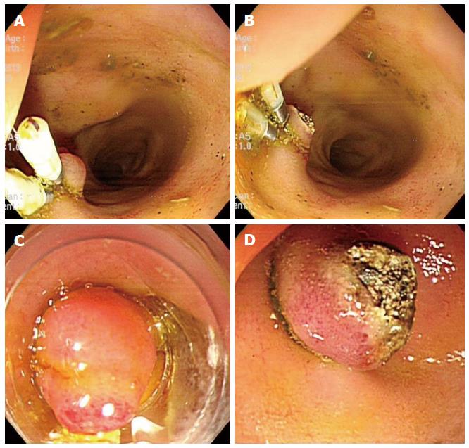
Figure 1 Experimental biliary dilatation model using endoclips and endoscopic band ligation with argon plasma coagulation.
A, B: Argon plasma coagulation (APC) was performed on the orifice of ampulla on the duodenal bulb following multiple rounds of hemoclipping on the ampulla of Vater; C, D: In an alternate model, endoscopic band ligation was performed on the ampulla of Vater, again followed by APC in the same manner.
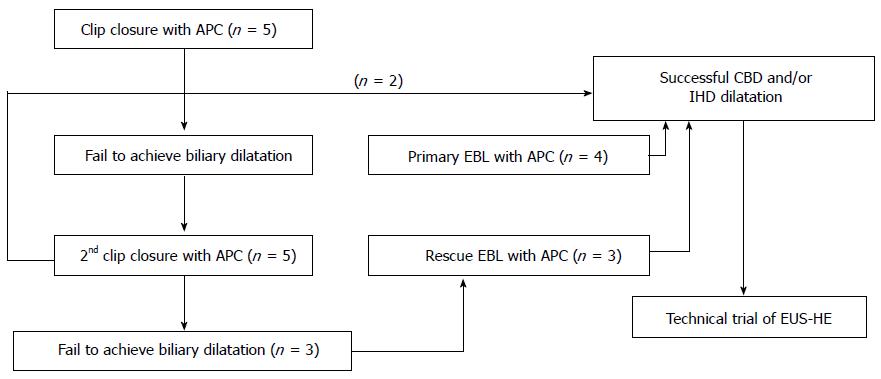
Figure 2 Flow of experimental endoscopic biliary dilatation using clip closure and/or endoscopic band ligation with argon plasma coagulation in a porcine model.
APC: Argon plasma coagulation; EBL: Endoscopic band ligation; IHD: Intrahepatic bile duct; CBD: Common bile duct; EUS-HE: Endoscopic ultrasound-guided hepaticoenterostomy.
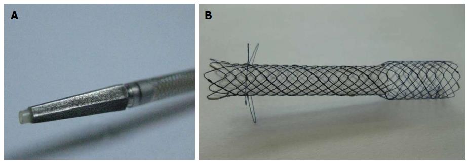
Figure 3 Gross finding of dedicated device for one-step endoscopic ultrasound-guided biliary drainage system.
A: The 7F delivery sheath and 3F catheter with 4F tapered pentagonal metal tip; B: Gross finding of the metallic stent. The uncovered proximal end of the stent (8 mm in diameter and 15 mm in length), the body and distal portions of the covered stent with a silicone membrane (6 mm in diameter and 35 or 45 mm in length), and the distal end with four flaps.
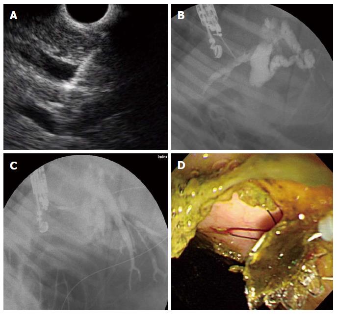
Figure 4 Endoscopic ultrasound-guided hepaticoenterostomy.
A: Linear endoscopic ultrasound (EUS) image shows a 19-G needle puncture targeting the intrahepatic bile duct (IHD; B: Fluoroscopic image reveals marked IHD and common bile duct dilatation with ampullary obstruction; C: After EUS-guided 19-G needle puncture of the IHD, the device for one-step endoscopic ultrasound-guided biliary drainage system was inserted and the stent released under the guidance of EUS and fluoroscopy; D: Following successful deployment of the stent, the bile passage was seen in the high body of stomach.
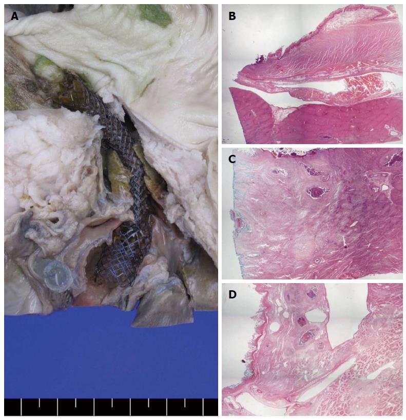
Figure 5 Gross and microscopic findings in the autopsy specimens.
A: Gross hepaticoenterostomy specimens showed that stent insertion along the far distal esophagus to the intrahepatic bile duct (IHD) via the liver parenchyma did not cause complications, such as stent dislocation or migration; B-D: Microscopic findings showed the distal esophagus (B), liver parenchyma (C), and IHD (D) sections adjacent to the inserted metallic stent. Surrounding mild inflammation and necrotic tissue were seen but without any other complications, such as abscess or perforation.
- Citation: Lee TH, Choi JH, Lee SS, Cho HD, Seo DW, Park SH, Lee SK, Kim MH, Park DH. A pilot proof-of-concept study of a modified device for one-step endoscopic ultrasound-guided biliary drainage in a new experimental biliary dilatation animal model. World J Gastroenterol 2014; 20(19): 5859-5866
- URL: https://www.wjgnet.com/1007-9327/full/v20/i19/5859.htm
- DOI: https://dx.doi.org/10.3748/wjg.v20.i19.5859


