Copyright
©2014 Baishideng Publishing Group Inc.
World J Gastroenterol. May 21, 2014; 20(19): 5685-5693
Published online May 21, 2014. doi: 10.3748/wjg.v20.i19.5685
Published online May 21, 2014. doi: 10.3748/wjg.v20.i19.5685
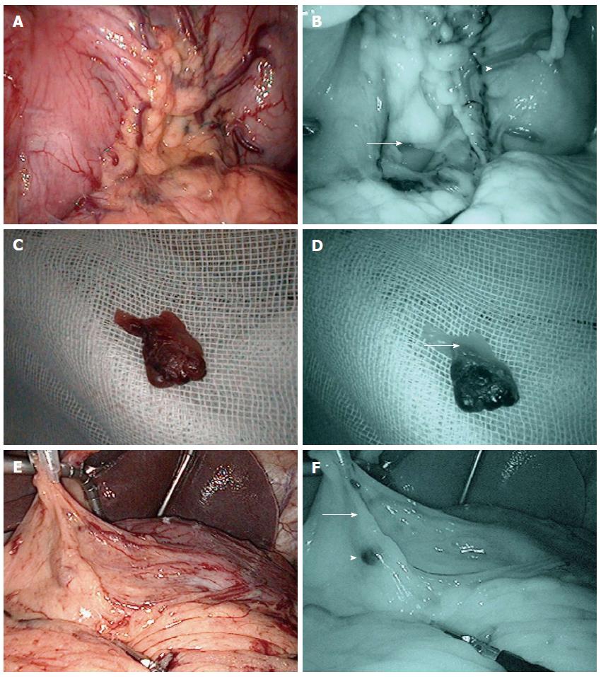
Figure 1 Laparoscopic observation around the left gastric vessels (LN No.
3,7). Lymph vessels and LN can be easily detected by IREE with Indocyanine Green (ICG). A: Ordinary light observation of lymph vessels around the left gastric artery; B: Infrared ray observation of lymph vessels around left gastric region (Lymph vessels: arrow; ICG positive node: arrowhead); C: Ordinary light observation of lymph vessels around the right epigastric artery; D: Infrared ray observation around the right epigastric artery shows first drainage lymph vessels, and sentinel lymph node (SLN) (arrow); E: Ordinary light observation of lymph vessels; F: Infrared ray observation of lymph vessels (first drainage lymph vessels: arrow; SLN: arrowhead).
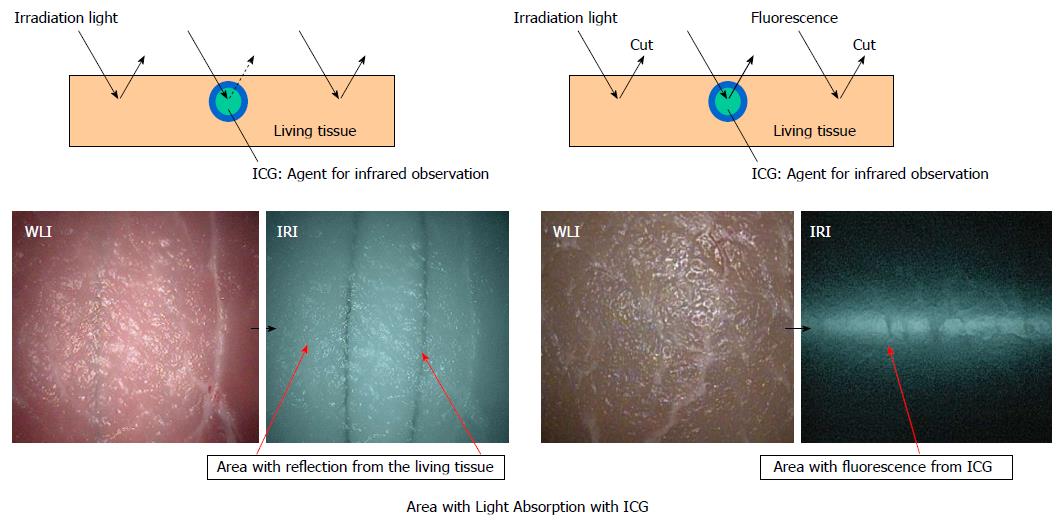
Figure 2 Mechanism of absorption with indocyanine green and fluorescence from indocyanine green.
Irradiated with light near the maximum absorption wavelength, the ICG-injected area in the tissue absorbs the light and becomes darker. In the other areas in the background, the light is reflected and those areas become brighter. ICG: Indocyanine Green.
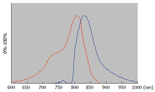
Figure 3 Wavelength of the indocyanine green.
Indocyanine green (ICG) has a maximum 805 nm of absorption wavelength. Absorption wavelength band of ICG: Red line; Fluorescence wavelength band of ICG: Blue line.
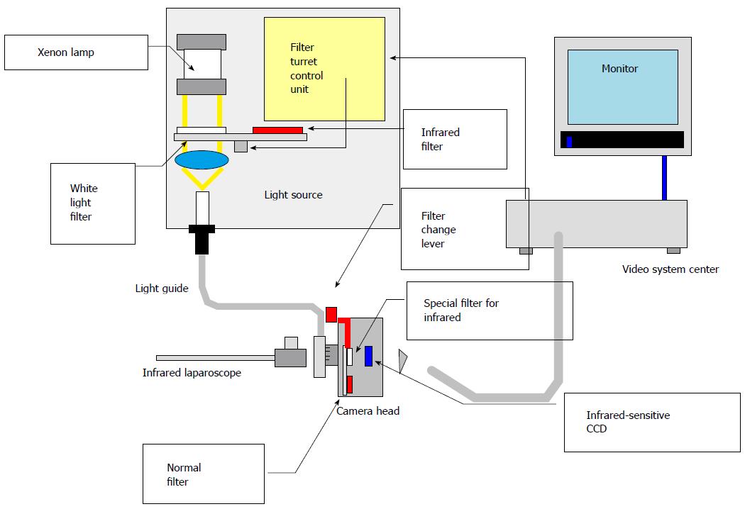
Figure 4 Technological overview of the new infrared observation system ordinary light and infrared ray laparoscopy system can be changed by a handle switch.
- Citation: Mitsumori N, Nimura H, Takahashi N, Kawamura M, Aoki H, Shida A, Omura N, Yanaga K. Sentinel lymph node navigation surgery for early stage gastric cancer. World J Gastroenterol 2014; 20(19): 5685-5693
- URL: https://www.wjgnet.com/1007-9327/full/v20/i19/5685.htm
- DOI: https://dx.doi.org/10.3748/wjg.v20.i19.5685


