Copyright
©2014 Baishideng Publishing Group Co.
World J Gastroenterol. May 14, 2014; 20(18): 5540-5547
Published online May 14, 2014. doi: 10.3748/wjg.v20.i18.5540
Published online May 14, 2014. doi: 10.3748/wjg.v20.i18.5540
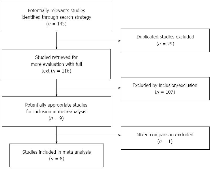
Figure 1 Process of article screening.
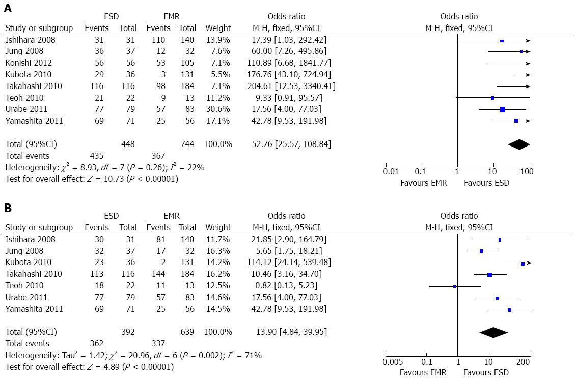
Figure 2 Comparison of en bloc resection (A) and curative rates (B) between endoscopic submucosal dissection and endoscopic mucosal resection.
ESD: Endoscopic submucosal dissection; EMR: Endoscopic mucosal resection.
Figure 3 Forest plot showing the procedural time for endoscopic submucosal dissection and endoscopic mucosal resection.
ESD: Endoscopic submucosal dissection; EMR: Endoscopic mucosal resection.
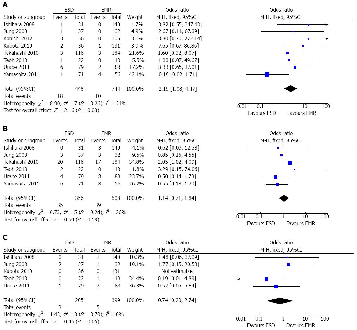
Figure 4 Incidence of perforation (A), stricture (B) and bleeding (C) between endoscopic submucosal dissection and endoscopic mucosal resection.
ESD: Endoscopic submucosal dissection; EMR: Endoscopic mucosal resection.
Figure 5 Comparison of recurrence rate between endoscopic submucosal dissection and endoscopic mucosal resection.
ESD: Endoscopic submucosal dissection; EMR: Endoscopic mucosal resection.
-
Citation: Guo HM, Zhang XQ, Chen M, Huang SL, Zou XP. Endoscopic submucosal dissection
vs endoscopic mucosal resection for superficial esophageal cancer. World J Gastroenterol 2014; 20(18): 5540-5547 - URL: https://www.wjgnet.com/1007-9327/full/v20/i18/5540.htm
- DOI: https://dx.doi.org/10.3748/wjg.v20.i18.5540


