Copyright
©2014 Baishideng Publishing Group Co.
World J Gastroenterol. May 7, 2014; 20(17): 5153-5156
Published online May 7, 2014. doi: 10.3748/wjg.v20.i17.5153
Published online May 7, 2014. doi: 10.3748/wjg.v20.i17.5153
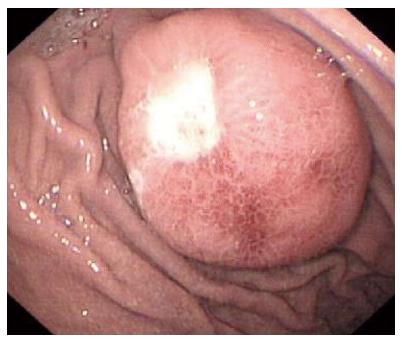
Figure 1 Endoscopic examination revealed a 3.
5 cm protruding and cauliflower-shaped mass with a shallow 1 cm central ulcer in the greater curvature of the stomach.
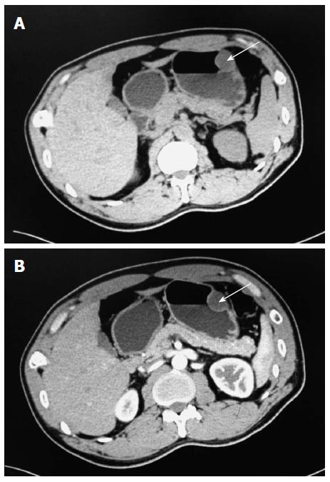
Figure 2 Radiological images of the abdomen.
A: Computed tomography (CT) showed a soft tissue mass-like lesion about 2.5 cm × 3.5 cm in the greater curvature of the stomach (arrow); B: Post-contrast abdominal CT shows the same area not representing the contrast-enhanced appearance (arrow).
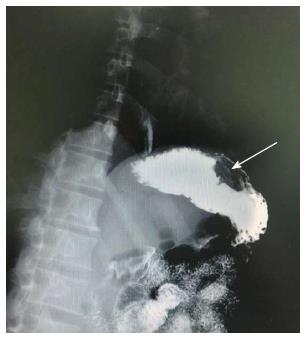
Figure 3 Upper gastrointestinal radiography showed a mass in the greater curvature of the stomach (arrow).
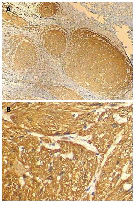
Figure 4 Histological examination of the gastric lesion.
A: Histological findings showing irregularly distorted, enlarged nerve bundles (× 40); B: Histopathological examination revealed nuclear and cytoplasmic reactivity with S-100 protein immunostain (× 400).
- Citation: Shi L, Liu FJ, Jia QH, Guan H, Lu ZJ. Solitary plexiform neurofibroma of the stomach: A case report. World J Gastroenterol 2014; 20(17): 5153-5156
- URL: https://www.wjgnet.com/1007-9327/full/v20/i17/5153.htm
- DOI: https://dx.doi.org/10.3748/wjg.v20.i17.5153


