Copyright
©2014 Baishideng Publishing Group Co.
World J Gastroenterol. May 7, 2014; 20(17): 5147-5152
Published online May 7, 2014. doi: 10.3748/wjg.v20.i17.5147
Published online May 7, 2014. doi: 10.3748/wjg.v20.i17.5147
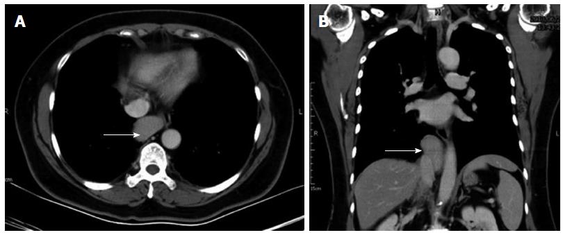
Figure 1 Computed tomography of the patient’s chest showing a well-circumscribed soft-tissue mass (white arrow), approximately 4.
35 cm × 2.5 cm × 6.14 cm in size, over the middle mediastinum, with mild compression on the esophagus. A: Sagittal view; B: Transverse view.
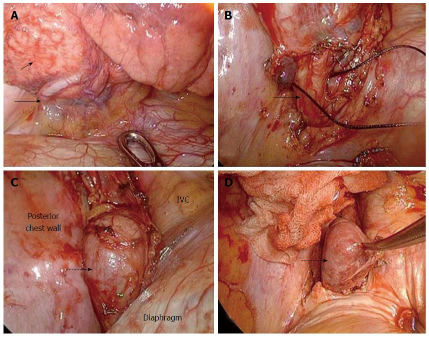
Figure 2 Intraoperative picture.
Intraoperative picture showing the exposure of the abnormal lung with hypervascularity at the posterior basal segment (A) (short arrow) and aberrant vessels (B) (long arrow) passing through diaphragm. The mass (C) (dotted arrow), covered by the sac, abutted the inferior vena cava (IVC), posterior chest wall and diaphragm. After dissecting the covering sac, a herniated liver (D) (dotted arrow) was impressed.
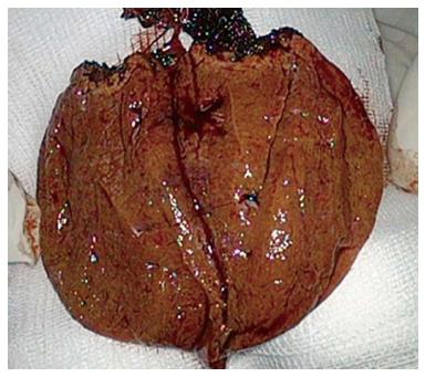
Figure 3 Cutting surface of the resected specimen showed liver tissue with cirrhotic changes.
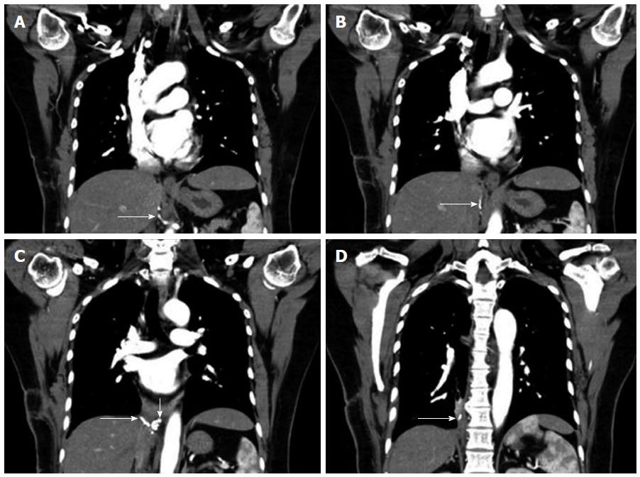
Figure 4 Postoperative computed tomography angiography confirmed the cutting end of the aberrant vessels.
A-D: Showed the trend of the aberrant vessel. The remnant aberrant vessel from abdominal aorta (long arrow), engorged aberrant vessels (short arrow).
- Citation: Chen YY, Huang TW, Chang H, Hsu HH, Lee SC. Intrathoracic caudate lobe of the liver: A case report and literature review. World J Gastroenterol 2014; 20(17): 5147-5152
- URL: https://www.wjgnet.com/1007-9327/full/v20/i17/5147.htm
- DOI: https://dx.doi.org/10.3748/wjg.v20.i17.5147


