Copyright
©2014 Baishideng Publishing Group Co.
World J Gastroenterol. Apr 21, 2014; 20(15): 4353-4361
Published online Apr 21, 2014. doi: 10.3748/wjg.v20.i15.4353
Published online Apr 21, 2014. doi: 10.3748/wjg.v20.i15.4353
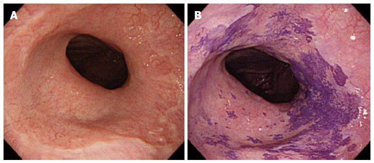
Figure 1 Barrett’s esophagus stained by crystal violet.
A: Regular observation of Barrett’s esophagus; B: Staining with crystal violet in the same region.
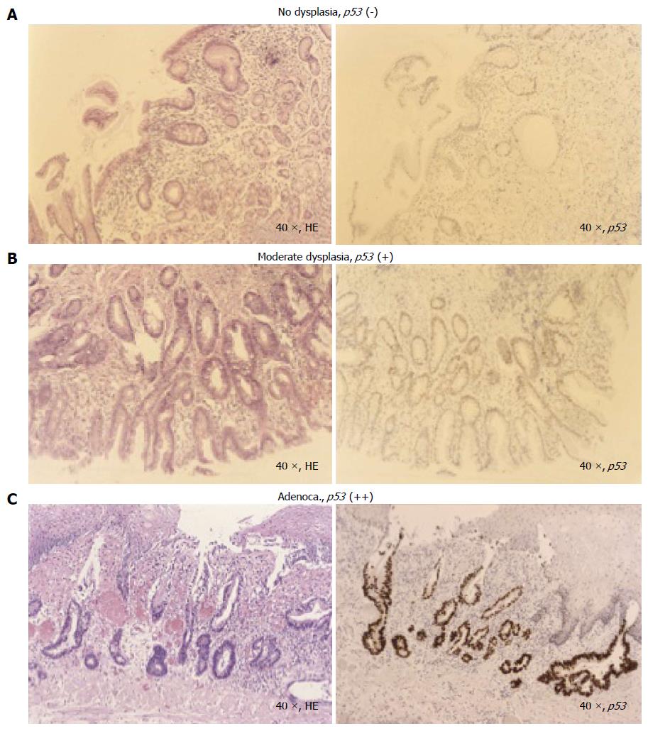
Figure 2 Immunostaining of p53.
The upper panel shows hematoxylin and eosin staining and the lower panel shows immunostaining of p53 using the identical sample. A: (-), no p53 expression; B: (+), moderate p53 expression characterized by positive nuclear staining in 5%-10% of cells; C: (++), high p53 expression characterized by positive nuclear staining in > 10% of cells.
- Citation: Fujita M, Nakamura Y, Kasashima S, Furukawa M, Misaka R, Nagahara H. Risk factors associated with Barrett’s epithelial dysplasia. World J Gastroenterol 2014; 20(15): 4353-4361
- URL: https://www.wjgnet.com/1007-9327/full/v20/i15/4353.htm
- DOI: https://dx.doi.org/10.3748/wjg.v20.i15.4353


