Copyright
©2014 Baishideng Publishing Group Co.
World J Gastroenterol. Apr 21, 2014; 20(15): 4160-4166
Published online Apr 21, 2014. doi: 10.3748/wjg.v20.i15.4160
Published online Apr 21, 2014. doi: 10.3748/wjg.v20.i15.4160
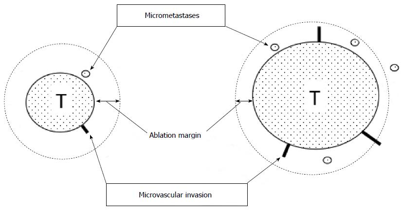
Figure 1 Ablation margin and micrometastases/microvascular invasion.
Radiofrequency ablation (RFA) therapy is required to ablate the main tumor and its surrounding liver tissues involving micrometastases and microvascular invasion. However, as the tumor get bigger, micrometastases and microcascular invasion frequently occur. Unablated lesions lead to local recurrences after RFA. T: Tumor.
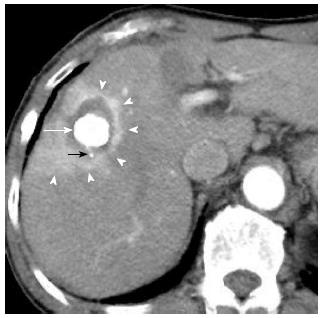
Figure 2 A 80-year-old woman with 2.
5 cm hepatocellular carcinoma after radiofrequency ablation combined with transcatheter arterial chemoembolization. Early-phase dynamic computed tomography shows a high-density center indicating Lipiodol deposition in hepatocellular carcinoma (white arrow) and a surrounding low-density zone indicating radiofrequency ablation-induced coagulation necrosis of the liver. A microsatellite (black arrow) was depicted as a high-density spot in the low-density zone. Therefore, this ablation therapy achieved complete necrosis of chief tumor and micrometastasis. Moreover, hyperemia surrounding the ablated lesion is depicted as peripheral rim enhancement (arrowheads).
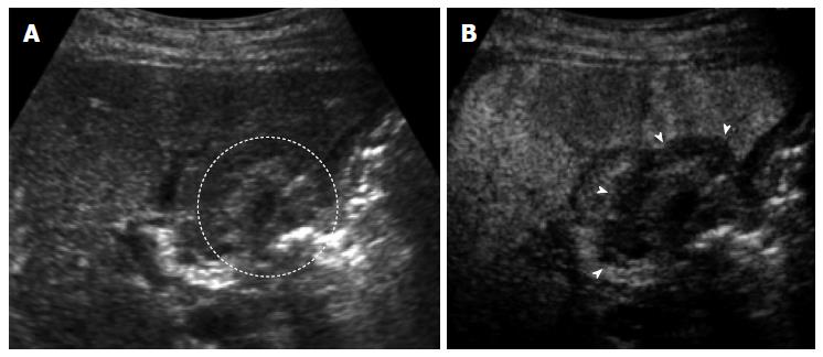
Figure 3 A 70-year-old man with 1.
5 cm hepatocellular carcinoma after radiofrequency ablation. A: The ablated tumor is depicted as hyper echoic lesion (circle) on B-mode ultrasound (US). However, the boundary between ablated area and unablated liver tissue could not be identified clearly; B: Contrast-enhanced US using Sonazoid shows the defect (arrows) in the Kupffer phase. The ablation margin is shown as low echoic zone surrounding the ablated tumor.
- Citation: Minami Y, Nishida N, Kudo M. Therapeutic response assessment of RFA for HCC: Contrast-enhanced US, CT and MRI. World J Gastroenterol 2014; 20(15): 4160-4166
- URL: https://www.wjgnet.com/1007-9327/full/v20/i15/4160.htm
- DOI: https://dx.doi.org/10.3748/wjg.v20.i15.4160


