Copyright
©2014 Baishideng Publishing Group Co.
World J Gastroenterol. Apr 14, 2014; 20(14): 4011-4016
Published online Apr 14, 2014. doi: 10.3748/wjg.v20.i14.4011
Published online Apr 14, 2014. doi: 10.3748/wjg.v20.i14.4011
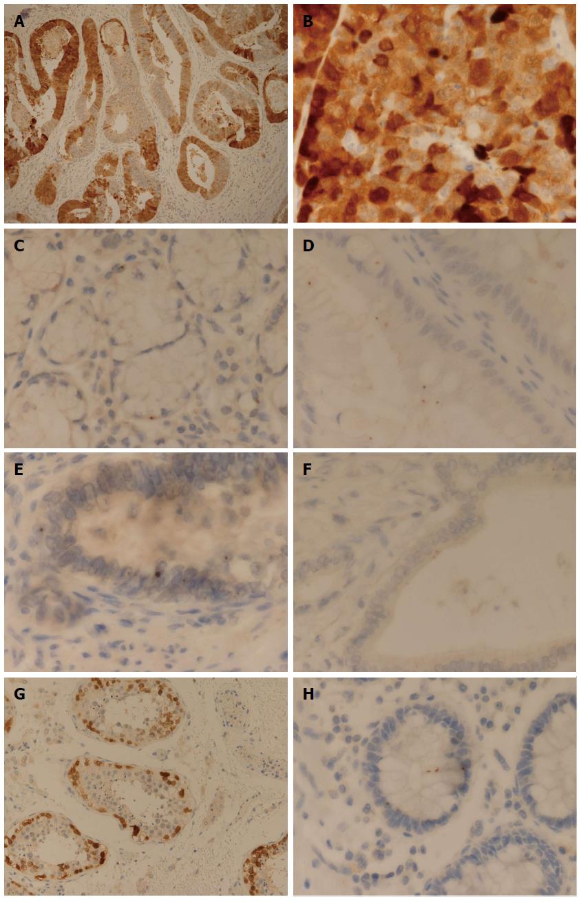
Figure 1 Immunohistochemistry images.
A: Esophageal adenocarcinoma (EAC) demonstrating diffuse, heterogeneous and mosaic-type expression for NY-ESO-1 (immunohistochemistry, IHC × 10); B: EAC showing a diffuse and granular pattern of cytoplasmic NY-ESO-1 expression (IHC × 40); C: Esophageal submucosal gland demonstrating dot-type expression for NY-ESO-1 (IHC × 60); D: Barrett’s esophagus of intestinal-type showing apical cytoplasmic dot-type expression for NY-ESO-1 (IHC × 60); E: EAC showing nuclear dot-type expression for NY-ESO-1 (IHC × 60); F: Well differentiated EAC with apical cytoplasmic dot-type expression for NY-ESO-1 (IHC × 60); G: Positive control (testis) demonstrating diffuse NY-ESO-1 expression which is restricted to primitive germ cells (IHC × 20); H: Negative control (colon) with dot-type pattern at nuclear, paranuclear and cytoplasmic locations, involving a basal crypt. Two of the cytoplasmic dots show alignment and apparent relationship to the paranuclear dot (IHC × 60).
- Citation: Hayes SJ, Hng KN, Clark P, Thistlethwaite F, Hawkins RE, Ang Y. Immunohistochemical assessment of NY-ESO-1 expression in esophageal adenocarcinoma resection specimens. World J Gastroenterol 2014; 20(14): 4011-4016
- URL: https://www.wjgnet.com/1007-9327/full/v20/i14/4011.htm
- DOI: https://dx.doi.org/10.3748/wjg.v20.i14.4011


