Copyright
©2014 Baishideng Publishing Group Co.
World J Gastroenterol. Apr 7, 2014; 20(13): 3597-3608
Published online Apr 7, 2014. doi: 10.3748/wjg.v20.i13.3597
Published online Apr 7, 2014. doi: 10.3748/wjg.v20.i13.3597
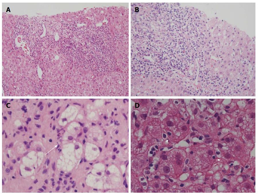
Figure 1 Histological features of primary biliary cirrhosis with interface hepatitis and autoimmune hepatitis.
A: Moderate interface hepatitis (score 3) in autoimmune hepatitis (AIH); B: Moderate interface hepatitis (score 3) in primary biliary cirrhosis (PBC); C: Rosette formation; regenerative hepatocytes arranged around a bile canaliculus (arrow), found in AIH; D: Emperipolesis, the engulfment of lymphocytes within hepatocytes (arrowheads), was found in areas of interface hepatitis. Hematoxylin and eosin stain, original magnifications × 200 ( A, B); × 600 (C, D).
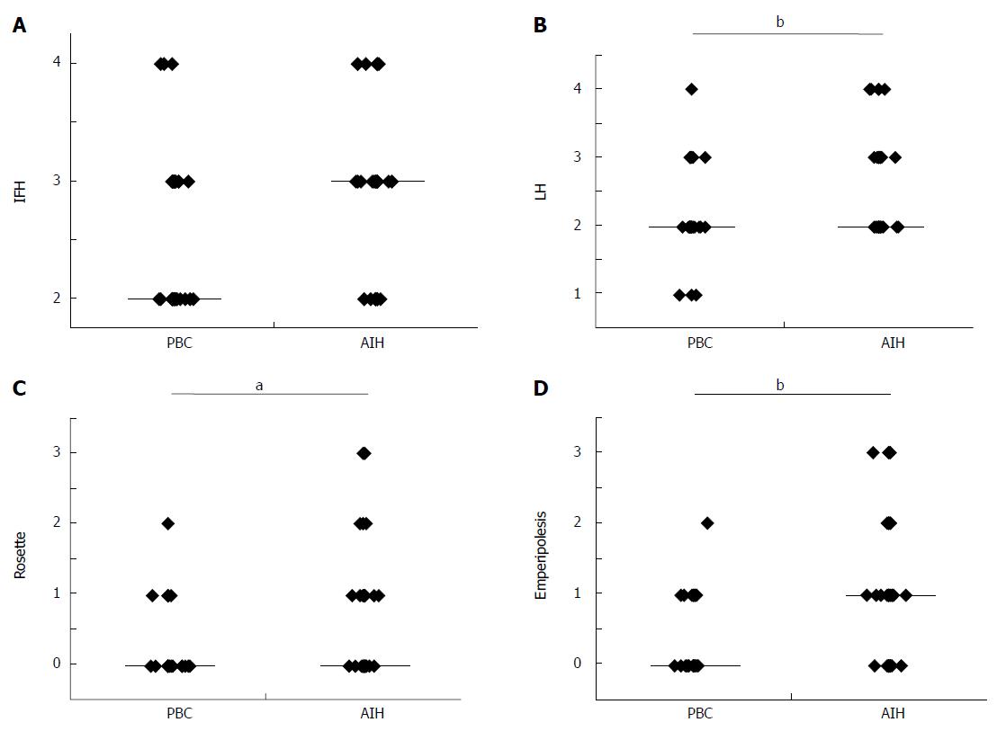
Figure 2 Comparison of necroinflammation in autoimmune hepatitis and primary biliary cirrhosis with interface hepatitis.
A: The scores of interface hepatitis (IFH) were not significantly different between primary biliary cirrhosis (PBC) (2.49 ± 0.64) and AIH (2.74 ± 0.66) (P = 0.0599) because PBC cases with IFH were chosen for this comparative study with autoimmune hepatitis (AIH); B: The scores of lobular hepatitis (LH) were higher in AIH (2.58 ± 0.70) than in PBC (2.22 ± 0.61) (P = 0.0003); C: The scores of hepatitic rosette formation were higher in AIH (0.55 ± 0.83) than in PBC (0.17 ± 0.44) (P = 0.0134); D: The scores of emperipolesis were higher in AIH (1.00 ± 0.96) than in PBC (0.32 ± 0.52) (P = 0.0003). Horizontal bars of the graph show the median scores; aP < 0.05 vs control, bP < 0.01 vs control in the Mann-Whitney test.
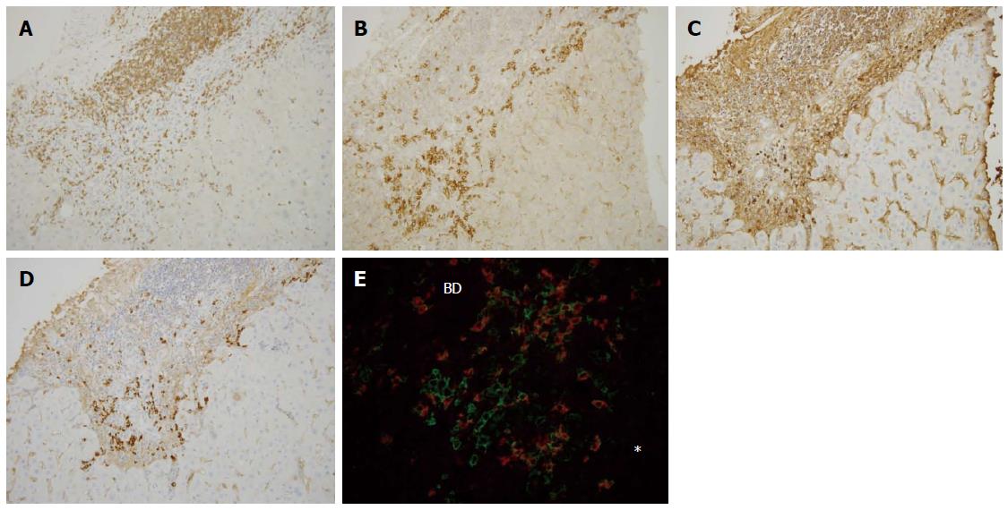
Figure 3 Immunohistochemical findings of primary biliary cirrhosis with interface hepatitis.
Many CD3+ cells were found within the portal tract and at the interface (A), whereas CD38+ cells heavily infiltrated the interface (B), IgG+ cells were mainly found in the portal tract and also at the interface (C), and IgM+ cell infiltration was predominant at the interface (D). Double staining for IgM (red) and CD38 (green) (E) showed that IgM-CD38 double positive cells, IgM-producing plasma cells, were frequently seen in the periportal area (star) (original magnifications: × 200). IgM+ plasma cells were also found around the intralobular bile duct. Asterisk: Hepatic lobule, BD: Bile duct.
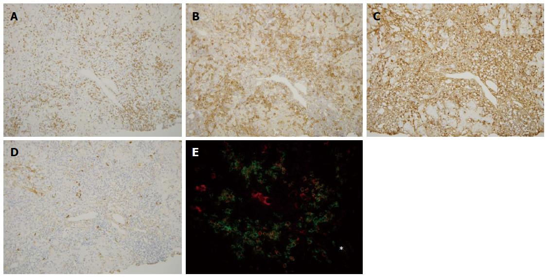
Figure 4 Immunohistochemical findings of autoimmune hepatitis.
CD3+ cells were found within the portal tract and in the periportal area (A), whereas CD38+ cells infiltrated prominently in the periportal area (B). IgG+ cells (C) and IgM+ cells (D) were also found in the periportal area, and IgG+ cell infiltration was predominant in the majority of autoimmune hepatitis cases. The distribution of these cells was similar to primary biliary cirrhosis (Figure 3); E: Double staining for IgG (red) and CD38 (green) showed that IgG-CD38 double positive cells, which may be IgG-producing plasma cells, were frequently seen in the periportal area (star). Original magnifications: × 200. Asterisk: Hepatic lobule.
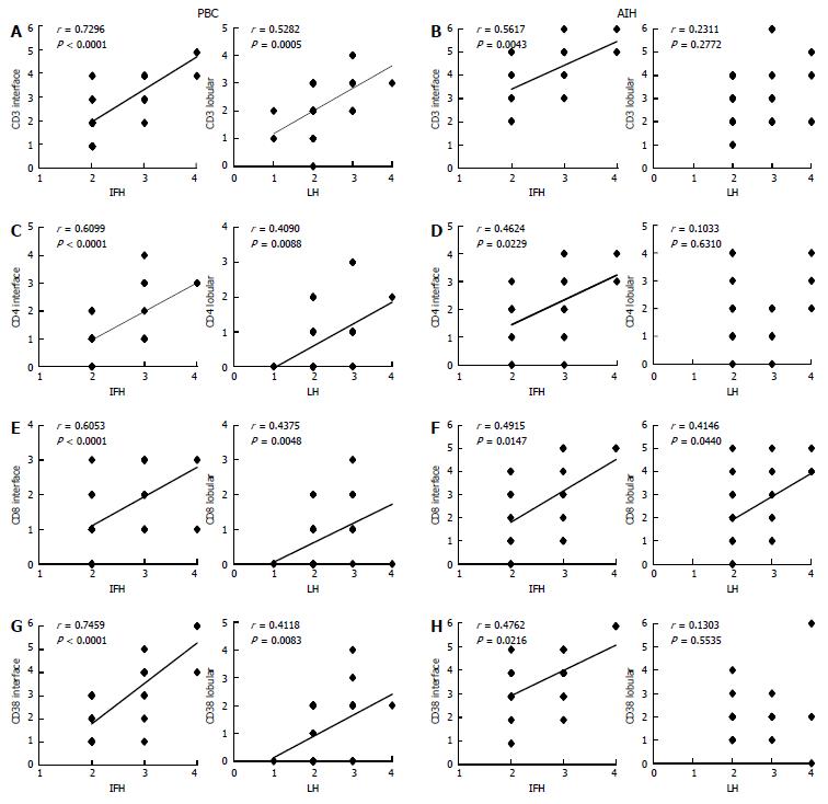
Figure 5 Correlation between scores for necroinflammation and infiltrating mononuclear cells positive for CD3, CD4, CD8, and CD38 in primary biliary cirrhosis with interface hepatitis and autoimmune hepatitis.
A: In primary biliary cirrhosis (PBC), the degree of CD3+ mononuclear cell infiltration and the scores of interface hepatitis and lobular hepatitis showed positive correlations; B: In autoimmune hepatitis (AIH), the degree of CD3+ mononuclear cell infiltration and the scores of interface hepatitis showed positive correlation, whereas that of CD3+ mononuclear cell infiltration and lobular hepatitis failed to correlate positively; C: In PBC, the degree of CD4+ mononuclear cell infiltration and the scores of interface hepatitis and lobular hepatitis showed positive correlations; D: In AIH, the degree of CD4+ mononuclear cell infiltration and the scores of interface hepatitis showed a positive correlation, whereas that of CD4+ mononuclear cell infiltration and lobular hepatitis failed to correlate positively; E: In PBC, the degree of CD8+ mononuclear cell infiltration and the scores of interface hepatitis and lobular hepatitis showed positive correlations; F: In AIH, the degree of CD8+ mononuclear cell infiltration and the scores of interface hepatitis and lobular hepatitis showed positive correlations; G: In PBC, the degree of CD38+ mononuclear cell infiltration and the scores of interface hepatitis and lobular hepatitis showed positive correlations; H: In AIH, the degree of CD38+ mononuclear cell infiltration and the scores of interface hepatitis showed positive correlation, whereas that of CD38+ mononuclear cell infiltration and lobular hepatitis failed to correlate positively. Spearman’s rank correlation test in A-H.
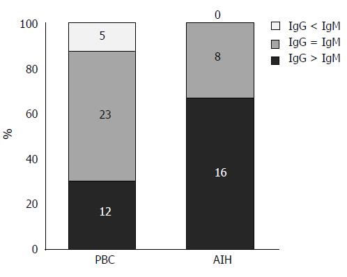
Figure 6 Classification of primary biliary cirrhosis with interface hepatitis and autoimmune hepatitis based on the predominance of the subclass of plasma cells infiltrating the interface.
The majority of autoimmune hepatitis (AIH) cases (16 cases) were IgG-predominant, whereas 8 cases were IgG/IgM-equal. There are no IgM-predominant AIH cases. In primary biliary cirrhosis (PBC), 23 cases were IgG/IgM-equal, whereas 12 cases were IgG-predominant and 5 cases were IgM-predominant.
- Citation: Kobayashi M, Kakuda Y, Harada K, Sato Y, Sasaki M, Ikeda H, Terada M, Mukai M, Kaneko S, Nakanuma Y. Clinicopathological study of primary biliary cirrhosis with interface hepatitis compared to autoimmune hepatitis. World J Gastroenterol 2014; 20(13): 3597-3608
- URL: https://www.wjgnet.com/1007-9327/full/v20/i13/3597.htm
- DOI: https://dx.doi.org/10.3748/wjg.v20.i13.3597


