Copyright
©2014 Baishideng Publishing Group Co.
World J Gastroenterol. Mar 28, 2014; 20(12): 3376-3382
Published online Mar 28, 2014. doi: 10.3748/wjg.v20.i12.3376
Published online Mar 28, 2014. doi: 10.3748/wjg.v20.i12.3376
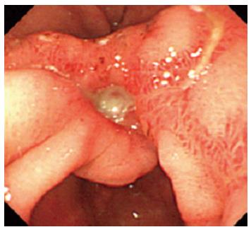
Figure 1 Initial gastrointestinal endoscopy shows gastric ulcers in the antrum.
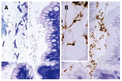
Figure 2 Histological findings in non-Helicobacter pylori helicobacter SH9 in gastric mucosa.
The organisms are present in small groups in gastric pits (A, B). The organisms display a tightly coiled spiral configuration and (B) are positive for the anti-Helicobacter pylori (H. pylori) antibody. (A) HE, original magnification, × 1000; (B) Immunostaining with anti-H. pylori antibody, original magnification, × 1000.
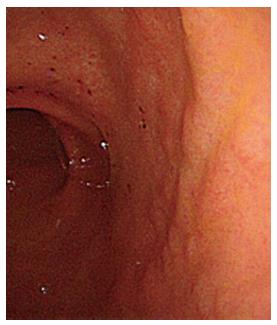
Figure 3 Gastrointestinal endoscopy after lansoprazole therapy shows erosion and a slightly coarse granular appearance in the antrum.
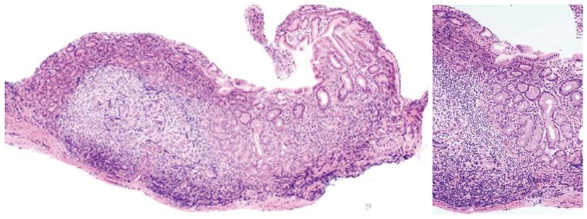
Figure 4 Histological findings of the lesser curvature of gastric antrum from a SH9 (Helicobacter heilmannii sensu stricto)-infected patient after the administration of lansoprazole for the treatment of gastric ulcers.
The gastric mucosa shows large lymphoid follicles with germinal centers without epithelial degeneration.
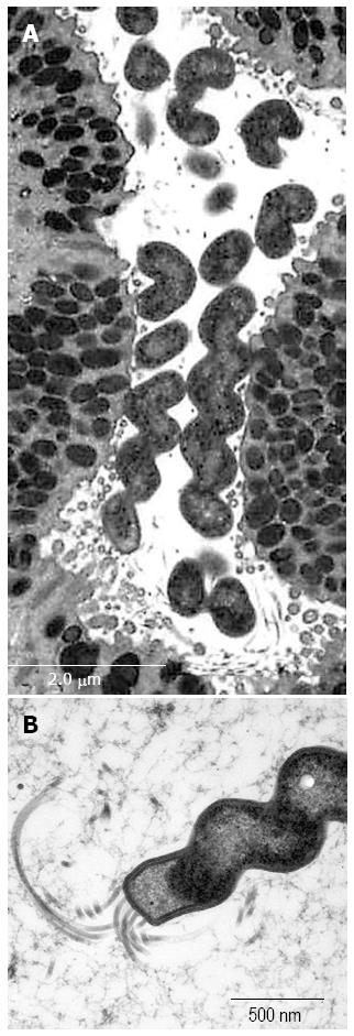
Figure 5 Ultrastructure of SH9 (Helicobacter heilmannii s.
s.) infected mouse gastric mucosa. The organisms showing tightly coiled spiral configuration (A) with flagella (B) are positioned on the apical surface of the surface mucous cells.
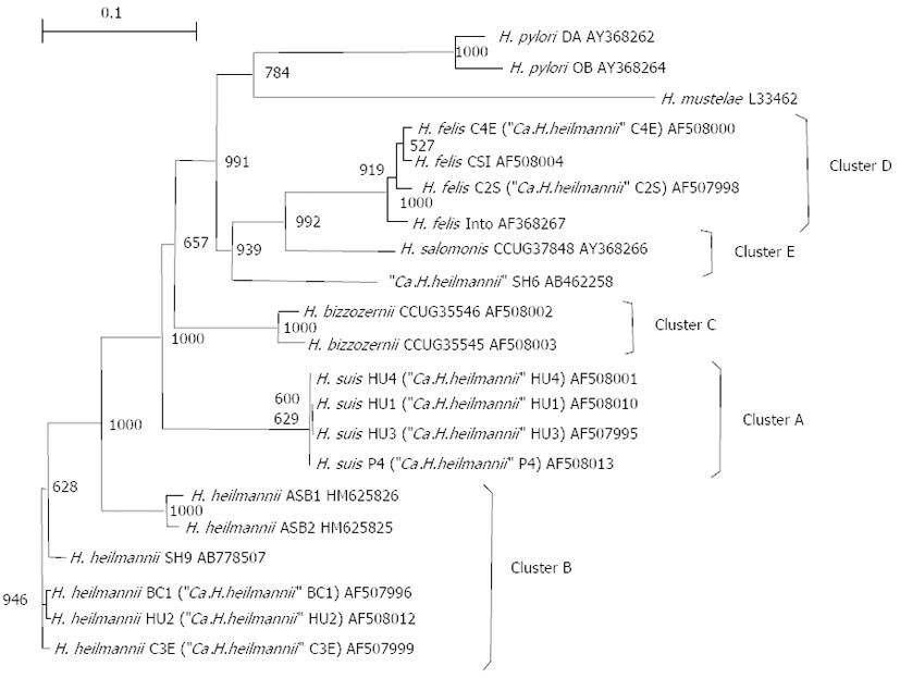
Figure 6 Phylogenetic tree based on partial ureA and ureB gene sequences, demonstrating the relationship between Helicobacter spp.
from animals and humans. Clusters A through E correspond to Helicobacter suis (H.suis), Helicobacter heilmannii sensu stricto (H. heilmannii s.s.), Helicobacter bizzozerronii (H. bizzozerronii), Helicobacter felis (H. felis), and Helicobacter salomonis (H. salomonis), as described by O'Rourke et al[11]. Ca.H.heilmannii: Candidatus H. heilmannii; H. pylori: Helicobacter pylori.
-
Citation: Matsumoto T, Kawakubo M, Akamatsu T, Koide N, Ogiwara N, Kubota S, Sugano M, Kawakami Y, Katsuyama T, Ota H.
Helicobacter heilmannii sensu stricto-related gastric ulcers: A case report. World J Gastroenterol 2014; 20(12): 3376-3382 - URL: https://www.wjgnet.com/1007-9327/full/v20/i12/3376.htm
- DOI: https://dx.doi.org/10.3748/wjg.v20.i12.3376


