Copyright
©2014 Baishideng Publishing Group Co.
World J Gastroenterol. Mar 21, 2014; 20(11): 3018-3024
Published online Mar 21, 2014. doi: 10.3748/wjg.v20.i11.3018
Published online Mar 21, 2014. doi: 10.3748/wjg.v20.i11.3018
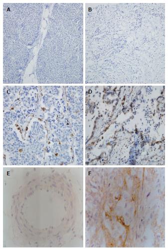
Figure 1 Representative immunohistochemistry results of midkine and syndecan-3 (× 200).
Paraffin sections were immunostained as described in Materials and Methods. A: Normal tissue showing negative midkine (MK) expression; B: Tumor tissue showing negative MK expression; C: Tumor tissue showing moderate MK expression; D: Tumor tissue showing intense MK expression; E: Negative syndecan-3 expression in cancer; F: Positive syndecan-3 expression in the perineurium of nerves in pancreatic cancer.
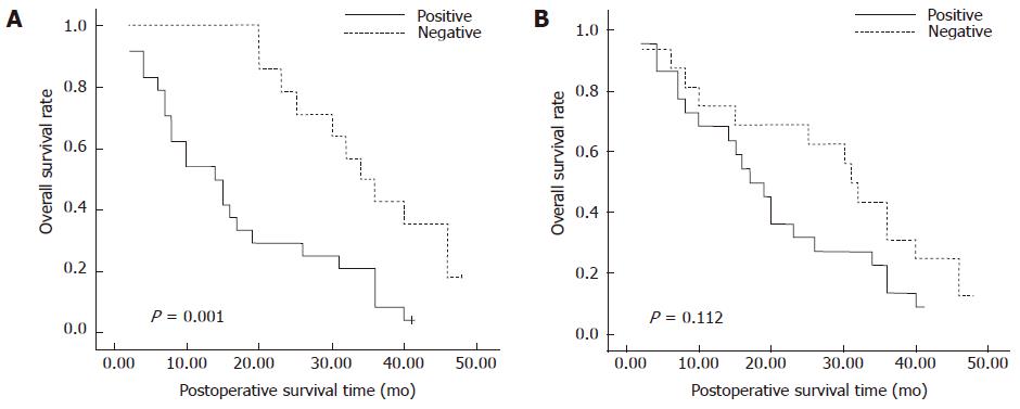
Figure 2 Kaplan-Meier analysis of overall postoperative survival curves in pancreatic cancer cases according to their immunohistochemical staining for midkine (A) and syndecan-3 (B).
- Citation: Yao J, Li WY, Li SG, Feng XS, Gao SG. Midkine promotes perineural invasion in human pancreatic cancer. World J Gastroenterol 2014; 20(11): 3018-3024
- URL: https://www.wjgnet.com/1007-9327/full/v20/i11/3018.htm
- DOI: https://dx.doi.org/10.3748/wjg.v20.i11.3018


