Copyright
©2014 Baishideng Publishing Group Co.
World J Gastroenterol. Jan 7, 2014; 20(1): 6-21
Published online Jan 7, 2014. doi: 10.3748/wjg.v20.i1.6
Published online Jan 7, 2014. doi: 10.3748/wjg.v20.i1.6
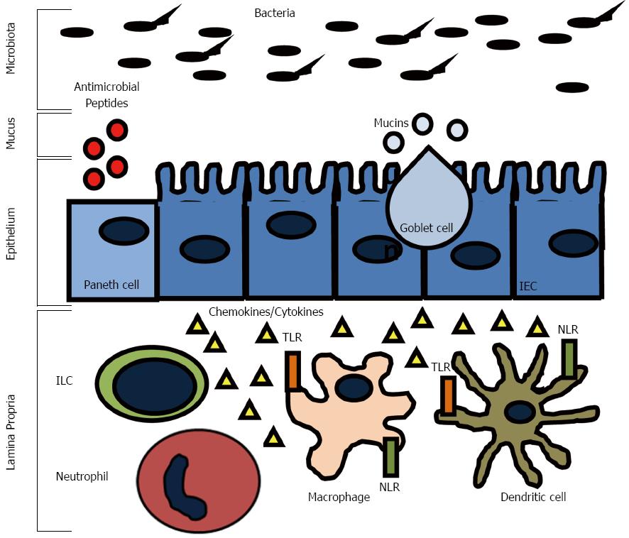
Figure 1 Innate immune system responses in the gut.
The intestinal epithelial barrier is equipped with several layers of defense mechanisms to limit luminal antigen translocation. Goblet cells, Paneth cells, and enterocytes secrete mucins and antimicrobial peptides that assemble into a mucus layer. Innate immune system cells, such as macrophages and dendritic cells, can sense invading bacteria through extracellular and intracellular pattern recognition receptors (Toll-like receptors-TRLs and NOD-like receptors-NLRs) and initiate rapid inflammatory reponses mediated by the secretion of cytokines and chemokines. Innate lymphoid cells (ILCs) are also found in the lamina propria where they contribute to cytokine production and inflammatory cell recruitment.
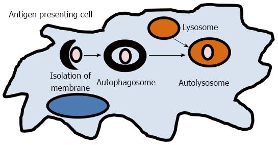
Figure 2 Autophagy.
A small volume of cytoplasm is enclosed by the autophagic isolation membrane, which forms the autophagosome. The outer membrane of the autophagosome then fuses with the lysosome where the cytoplasm derived materials are degraded.
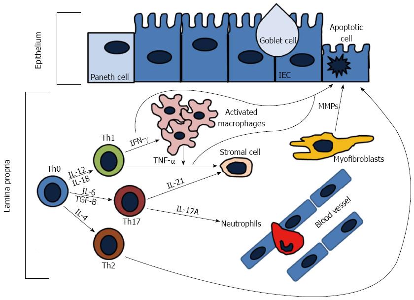
Figure 3 Adaptive immune responses in the gut.
During active inflammation, naïve T-cells (Th0) differentiate into T helper cell types (Th1, Th2, Th17) under stimulation of different cytokines. Th1 cells produce interferon (IFN)-γ and tumor necrosis factor (TNF)-α. IFN-γ activated tissue macrophages to produce additional TNF-α, which causes epithelial cell apoptosis and differentiation of stromal cells into myofibroblasts. Activated myofibroblasts produce metalloproteinases (MMPs) that cause tissue degredation. Th2 cells produce interleukin (IL)-13 that can increase intestinal permeability and induce epithelial apoptosis. Th17 cells release IL-17A, which plays a role in recruiting neutrophils to sites of active inflammation, and IL-21 that also induces MMP production that contributes to extracellular matrix degredation.
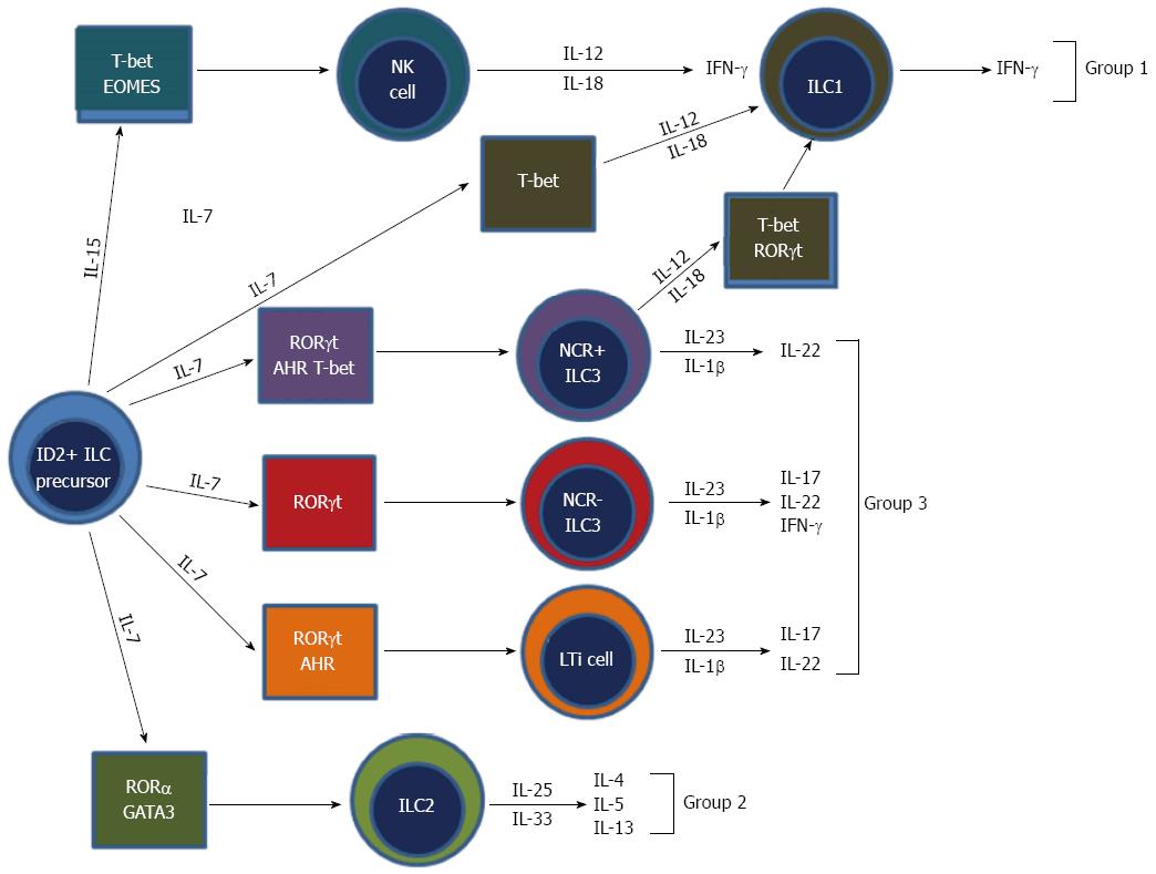
Figure 4 Development and classification of innate lymphoid cells[82].
Innate lymphoid cells (ILCs) all derive from an ID2 positive progenitor cell. Group 1 ILCs make interferon (IFN)-γ. Group 2 ILCs produce IL-5 and interleukin (IL)-13. Group 3 ILCs produce IL-17, IL-22, and IFN-γ. NK require IL-15, whereas all other ILCs require IL-7 for development. Group 2 ILCs depend on transcription factors GATA3 and ROR for development. Group 3 ILCs require RORt for development. Also, subsets of group 3 ILCs require additional transcription factors, such as aryl hydrocarbon receptor (AHR) for development. NK cells, which are group 1 ILCs, require both T-bet and eomesodermin (EOMES). The mechanisms of ILC1 development are not fully elucidated, however are known to require transcription factor T-bet for development.
- Citation: Wallace KL, Zheng LB, Kanazawa Y, Shih DQ. Immunopathology of inflammatory bowel disease. World J Gastroenterol 2014; 20(1): 6-21
- URL: https://www.wjgnet.com/1007-9327/full/v20/i1/6.htm
- DOI: https://dx.doi.org/10.3748/wjg.v20.i1.6


