Copyright
©2014 Baishideng Publishing Group Co.
World J Gastroenterol. Jan 7, 2014; 20(1): 323-325
Published online Jan 7, 2014. doi: 10.3748/wjg.v20.i1.323
Published online Jan 7, 2014. doi: 10.3748/wjg.v20.i1.323
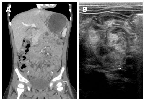
Figure 1 Imaging.
A: An abdominal computed tomography scan shows a large, nodular soft tissue mass occupying the pylorus and extending into the duodenum; B: Ultrasonography shows marked thickening of the mucosal and muscular layered lesion at the pylorus and duodenum.
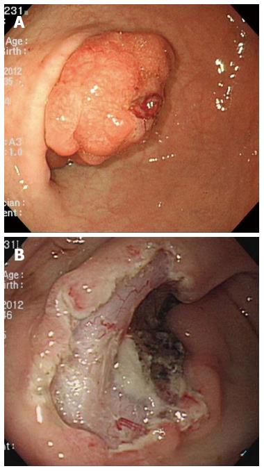
Figure 2 Endoscopic views.
A: Endoscopy shows a diffuse hyperemic and edematous giant lobulating mass from the pylorus to the duodenal inlet. B: The mass lifted easily and was resected in one piece endoscopically.
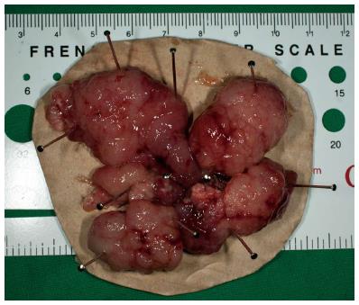
Figure 3 Retrieved pieces of the large polyp were reconstructed.
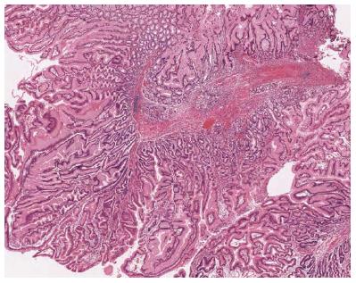
Figure 4 Photomicrograph of the polyp with thick-walled vessels and organized thick bundles of arborizing smooth muscle with a prominent basal glandular component.
- Citation: Jung EY, Choi SO, Cho KB, Kim ES, Park KS, Hwang JB. Successful endoscopic submucosal dissection of a giant polyp in a 21-month-old female. World J Gastroenterol 2014; 20(1): 323-325
- URL: https://www.wjgnet.com/1007-9327/full/v20/i1/323.htm
- DOI: https://dx.doi.org/10.3748/wjg.v20.i1.323


