Copyright
©The Author(s) 1996.
World J Gastroenterol. Dec 15, 1996; 2(4): 243-245
Published online Dec 15, 1996. doi: 10.3748/wjg.v2.i4.243
Published online Dec 15, 1996. doi: 10.3748/wjg.v2.i4.243
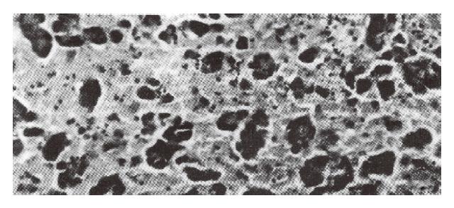
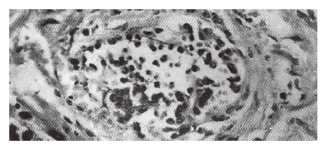
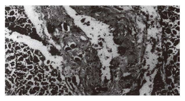
Figure 1 Hemorrhagic necrosis of the pancreas.
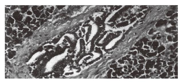
Figure 2 Shed epithelial cells and pseudoglandular formation.
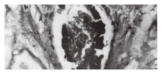
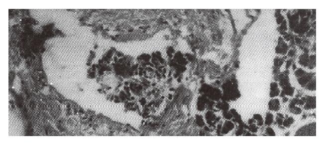
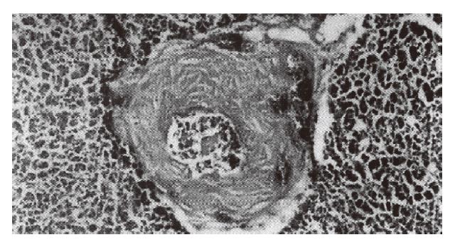
Figure 3 Massive blood in the pancreatic duct.
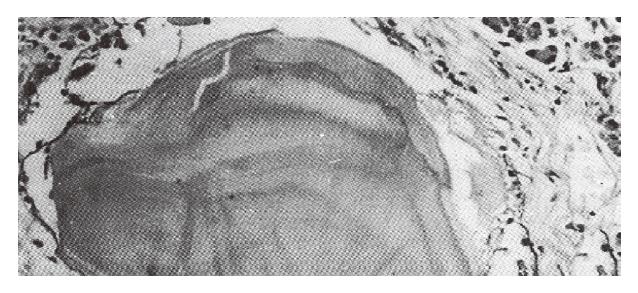
Figure 4 Eosinophilic material in the lumen of the pancreatic duct.
Figure 5 Eosinocytes in the lumen of the pancreatic duct.
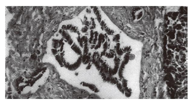
Figure 6 Papillary hyperplasia and neoplastic plug appearance.
Figure 7 Squamous metaplasia of the epithelial cells of the pancreatic duct.
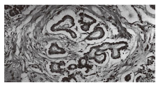
Figure 8 Shedding of the epithelium and intramural ductule formation in the wall of the pancreas.
Figure 9 Multiple intramural ductules lined with a layer of the epithelial cells with basement membrane.
Figure 10 As many as 6 intramural ductules.
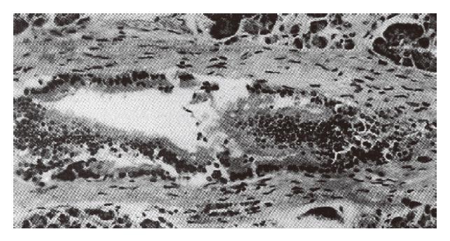
Figure 11 Intramural hemorrhage of the pancreatic duct.
- Citation: Hu SX, Tang RJ, Zuo DY, Chen PH. Histopathological study of pancreatic duct in acute fulminant pancreatitis. World J Gastroenterol 1996; 2(4): 243-245
- URL: https://www.wjgnet.com/1007-9327/full/v2/i4/243.htm
- DOI: https://dx.doi.org/10.3748/wjg.v2.i4.243


