Copyright
©The Author(s) 1996.
World J Gastroenterol. Dec 15, 1996; 2(4): 232-235
Published online Dec 15, 1996. doi: 10.3748/wjg.v2.i4.232
Published online Dec 15, 1996. doi: 10.3748/wjg.v2.i4.232
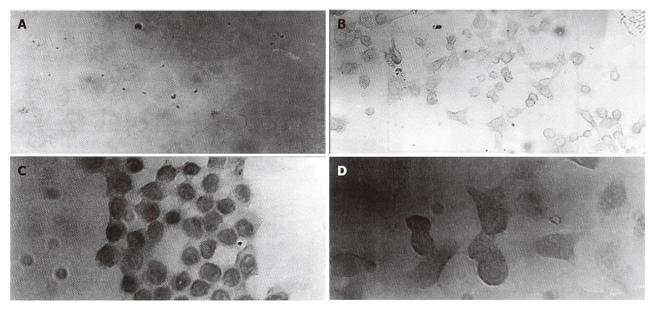
Figure 1 Demonstration of P-glycoprotein in HCT-8 (A and C) and HCTV2000 cells (B and D) by immunocytochemical staining with JSB-1 (A and B) and MRK-16 (C and D).
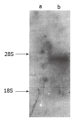
Figure 2 Northern hybridization.
Ten micrograms of total cellular RNA were loaded for each line. a. HCT-8 cell line; b. HCTV2000 cell line.
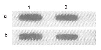
Figure 3 Slot blot hybridization.
Twenty micrograms (1) or 10 μg (2) DNA from HCT-8 cell line (a) and HCTV2000 cell line (b) were analyzed.
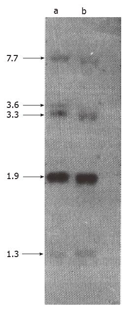
Figure 4 Southern hybridization of mdr1 gene.
Ten micrograms of DNA extracted from HCT-8 cell line (a) and HCTV2000 cell line (b) were digested with EcoR I and separated on a 0.7% agarose gel. The arrows indicate the position of hybridization.
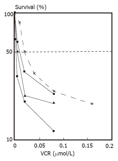
Figure 5 Effect of VRM on sensitivity of HCT-8 cells to VCR.
The doses of VRM were 0 (x), 2.5 (■), 5 (▲) and 10 (●) μg/mL.
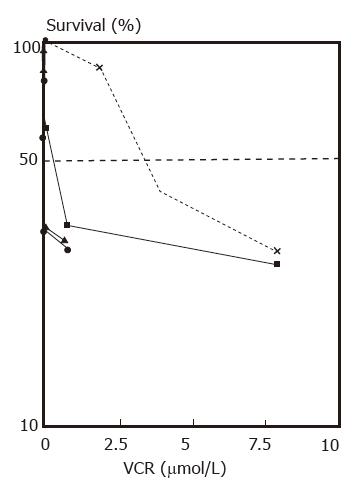
Figure 6 Effect of VRM o-n sensitivity of HCTV2000 cells to VCR.
The doses of VRM were o (x), 2.5 (■), 5 (▲) and 10 (●) μg/mL.
- Citation: Zhang XH, Li XT, Ji XJ, Zhu YJ. Preliminary study on mechanism of drug resistance in human colon cancer cell line HCTV2000. World J Gastroenterol 1996; 2(4): 232-235
- URL: https://www.wjgnet.com/1007-9327/full/v2/i4/232.htm
- DOI: https://dx.doi.org/10.3748/wjg.v2.i4.232


