Copyright
©The Author(s) 1996.
World J Gastroenterol. Sep 15, 1996; 2(3): 158-160
Published online Sep 15, 1996. doi: 10.3748/wjg.v2.i3.158
Published online Sep 15, 1996. doi: 10.3748/wjg.v2.i3.158
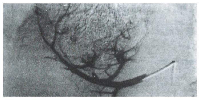
Figure 1 PHC treated by HAE with Lp MMC and GF powders.
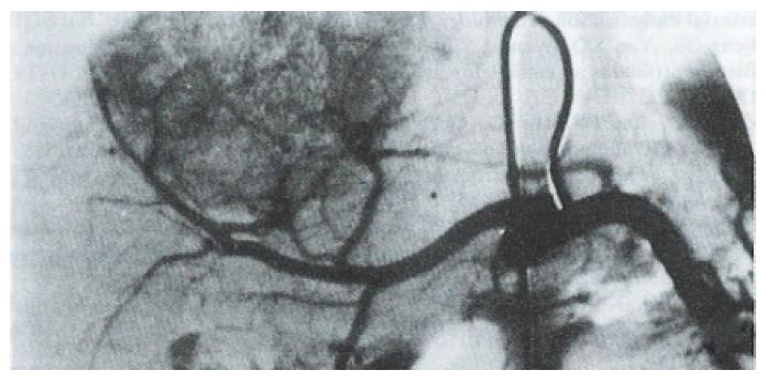
Figure 2 Three months after HAE, angiography of this patient showed tumor staining and slight shrinkage.
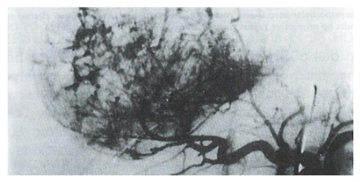
Figure 3 The right lobar massive PHC treated by HAE with Lp-MMC and BS powders.
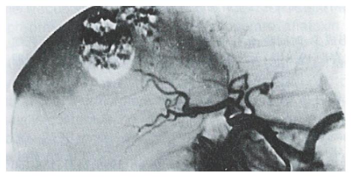
Figure 4 Ten months after HAE, angiography of this patient showed obvious tumor shrinkage, and no tumor vessels or collateral circulation.
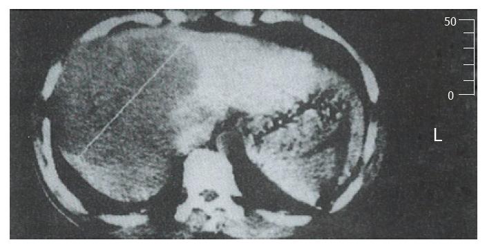
Figure 5 Prior to HAE with Lp MMC and BS powders, a CT scan of the massive PHC showed local low density in the right hepatic lobe.
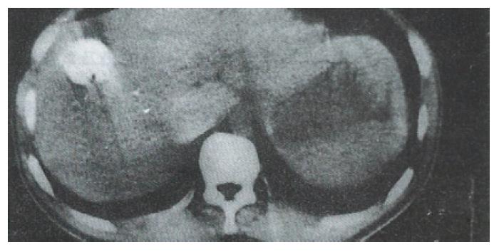
Figure 6 Nine months after HAE, a CT scan of this patient showed obvious tumor shrinkage and Lipiodol retention in the tumor area.
- Citation: Feng GS, Kramann B, Zheng CS, Zhou RM, Liang B, Zhang YF. Comparative study on the effects of hepatic arterial embolization with Bletilla striata or Gelfoam in treatment of primary hepatic carcinoma. World J Gastroenterol 1996; 2(3): 158-160
- URL: https://www.wjgnet.com/1007-9327/full/v2/i3/158.htm
- DOI: https://dx.doi.org/10.3748/wjg.v2.i3.158


