Copyright
©2013 Baishideng Publishing Group Co.
World J Gastroenterol. Mar 7, 2013; 19(9): 1494-1497
Published online Mar 7, 2013. doi: 10.3748/wjg.v19.i9.1494
Published online Mar 7, 2013. doi: 10.3748/wjg.v19.i9.1494
Figure 1 Colonoscopy and endoscopic ultrasonography results.
A, B: Colonoscopy showed an extensive hypervascular submucosal lesion, with tortuous submucosal veins and nodular mucosa; C: Endoscopic ultrasonography revealed an extensive anechoic mass with clear demarcation.
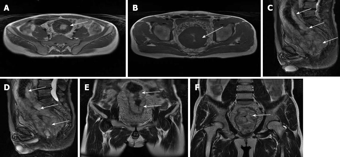
Figure 2 Magnetic resonance imaging showed a significant thickness of the rectal wall, extending to the distal edge of the anus, with a narrow lumen (arrows).
A, B: Horizontal plane imaging; C, D: Sagittal plane imaging; E, F: Coronal plane imaging.
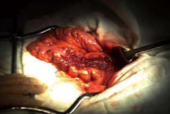
Figure 3 Diffuse colorectal proliferative lesions with no ascites or peritoneal dissemination were found during the operation.
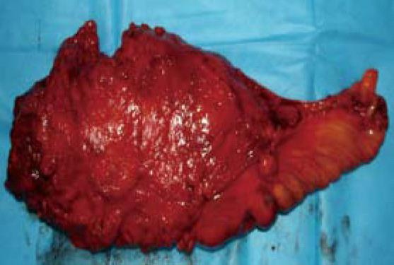
Figure 4 The rectal mass weighed 840 g and measured 20 cm × 8 cm × 8 cm, presenting as a cavernous, soft and compressible tumor.
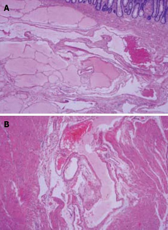
Figure 5 The tumor was composed of lymphatic and blood vessels mainly at the submucosa, occupied the entire wall (A), and extending into the surrounding fatty tissues (B).
- Citation: Chen G, Cui W, Ji XQ, Du JF. Diffuse hemolymphangioma of the rectum: A report of a rare case. World J Gastroenterol 2013; 19(9): 1494-1497
- URL: https://www.wjgnet.com/1007-9327/full/v19/i9/1494.htm
- DOI: https://dx.doi.org/10.3748/wjg.v19.i9.1494


