Copyright
©2013 Baishideng Publishing Group Co.
World J Gastroenterol. Dec 14, 2013; 19(46): 8793-8798
Published online Dec 14, 2013. doi: 10.3748/wjg.v19.i46.8793
Published online Dec 14, 2013. doi: 10.3748/wjg.v19.i46.8793
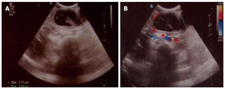
Figure 1 Abdominal ultrasonography demonstrated a solid cystic mass within the body of the pancreas.
A: Ultrasonography showed a solid cystic mass within the body of the pancreas; B: Ultrasonography showed that the mass was surrounded by blood vessels.
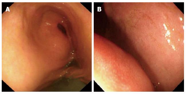
Figure 2 Gastroscopic imaging demonstrating fluid within the stomach and wall compression caused by an external mass.
A: Gastroscopy demonstrated intragastric fluid retention; B: Stomach wall compression by an external mass.
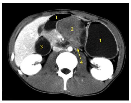
Figure 3 Abdominal computed tomography with contrast (venous phase) demonstrating a solid cystic mass invading the horizontal portion of the duodenum.
1: Stomach; 2: Pancreatic cystic mass; 3: Enlarged duodenum; 4: Horizontal duodenal portion invaded by the tumor.
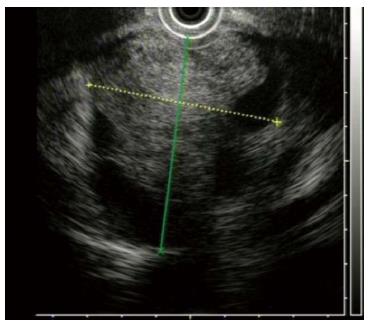
Figure 4 Endoscopic ultrasonography revealing a pancreatic cystic hypoechoic tumor located posterior to the stomach, with four cystic cavities, about 6.
7 cm × 5.5 cm in dimension.
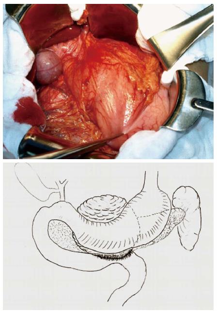
Figure 5 Exploratory laparotomy revealed a cystic mass of size 8.
6 cm × 6.0 cm within the pancreatic neck and body invading the horizontal duodenum.
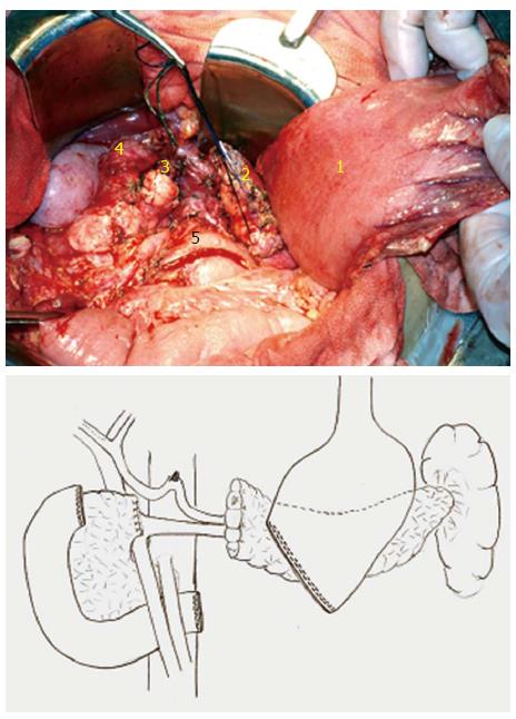
Figure 6 The central pancreas (containing the tumor), distal stomach and duodenal portion were all resected.
1: Stomach; 2: Left resected end of pancreas; 3: Right resected end of pancreas; 4: Upper resected end of duodenum; 5: Lower resected end of duodenum.
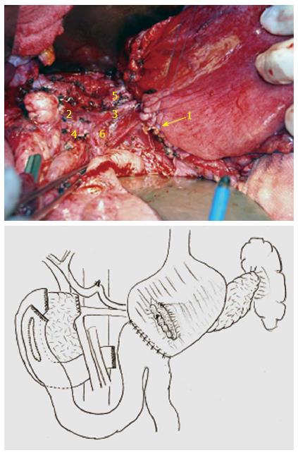
Figure 7 Reconstruction was performed via pancreaticogastrostomy, duodenojejunostomy (side to side) and gastrojejunostomy (Billroth II).
1: Pancreaticogastrostomy; 2: Portal vein; 3 Splenic vein; 4: Superior mesenteric vein; 5: Splenic artery; 6: Superior mesenteric artery.
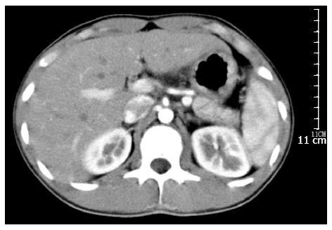
Figure 8 Contrast computed tomography examination (arterial phase) demonstrated no recurrence 18 mo after operation.
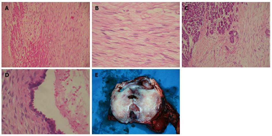
Figure 9 Pathological examination.
A: Pancreatic desmoid tumor with invasion to the duodenal wall; B: Proliferation of spindle-shaped stellate cells in fasciculate and storiform growth patterns within a myxoid intercellular matrix; C: Pancreatic infiltration by the tumor; D: The cystic area resulted from dilatation of entrapped excretory pancreatic ducts; E: Gross view of resected tumor and pancreas.
- Citation: Xu B, Zhu LH, Wu JG, Wang XF, Matro E, Ni JJ. Pancreatic solid cystic desmoid tumor: Case report and literature review. World J Gastroenterol 2013; 19(46): 8793-8798
- URL: https://www.wjgnet.com/1007-9327/full/v19/i46/8793.htm
- DOI: https://dx.doi.org/10.3748/wjg.v19.i46.8793


