Copyright
©2013 Baishideng Publishing Group Co.
World J Gastroenterol. Dec 14, 2013; 19(46): 8722-8730
Published online Dec 14, 2013. doi: 10.3748/wjg.v19.i46.8722
Published online Dec 14, 2013. doi: 10.3748/wjg.v19.i46.8722
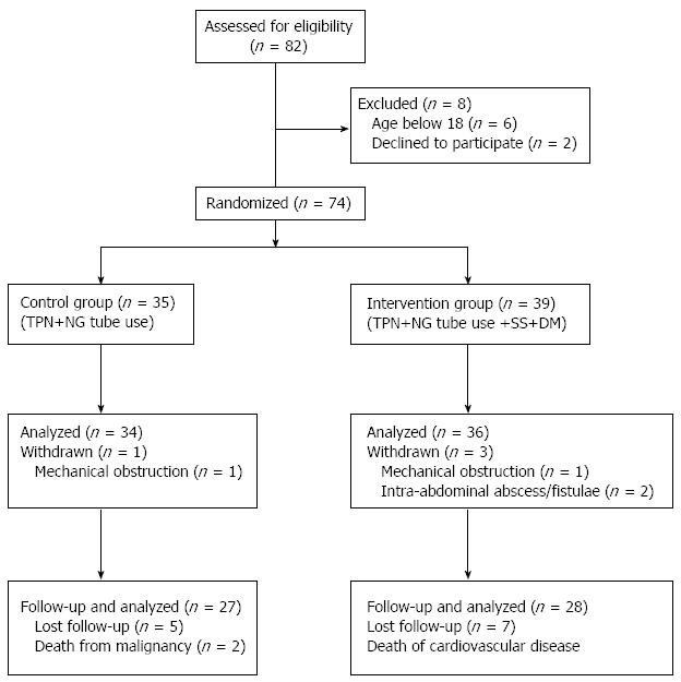
Figure 1 Flow chart of patient inclusion and follow-up.
TPN: Total parenteral nutrition; NG: Nasogastric; DM: Dexamethasone; SS: Somatostatin.
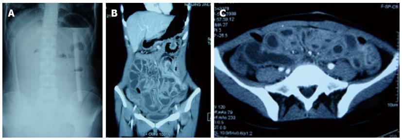
Figure 2 Typical radiographic and intra-operative finding in the last operation.
A: Typical upright radiograph of early postoperative small bowel obstruction with obliterative peritonitis after extensive adhesiolysis for abdominal cocoon, showing only mild air-fluid levels. No isolated small bowel loops were observed; B, C: Computed tomography scan reveals edematous small bowel filled-up with fluids. The border between the small bowel loops is not clear. No significant discrepancies in small bowel diameter and air-fluid levels were observed.
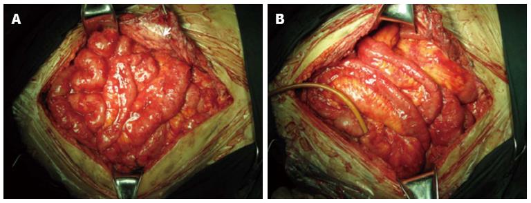
Figure 3 Intraoperative photo of a patient who developed early postoperative small bowel obstruction with obliterative peritonitis.
The 40-year-old male had a colostomy and patch-repair (arrow) of the abdominal defect after open abdomen due to trauma. He required closure of the stoma 6 months after colostomy. On operation, dense matted adhesions were found, especially beneath the patch, and full enterolysis resulted in extensive intestinal deserosalization (A). An intestinal splinting was performed (B). On postoperative day (POD) 10 the tube was removed, and he had a temporary return of bowel function but early postoperative small bowel obstruction with obliterative peritonitis on POD 15.
- Citation: Gong JF, Zhu WM, Yu WK, Li N, Li JS. Conservative treatment of early postoperative small bowel obstruction with obliterative peritonitis. World J Gastroenterol 2013; 19(46): 8722-8730
- URL: https://www.wjgnet.com/1007-9327/full/v19/i46/8722.htm
- DOI: https://dx.doi.org/10.3748/wjg.v19.i46.8722


