Copyright
©2013 Baishideng Publishing Group Co.
World J Gastroenterol. Dec 14, 2013; 19(46): 8562-8570
Published online Dec 14, 2013. doi: 10.3748/wjg.v19.i46.8562
Published online Dec 14, 2013. doi: 10.3748/wjg.v19.i46.8562
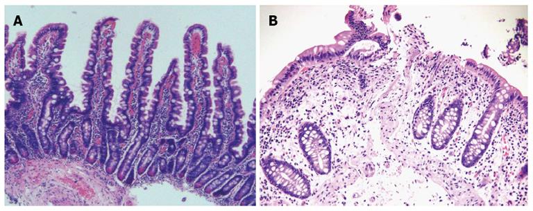
Figure 1 Histological appearance respectively.
A: Normal duodenal pattern; B: Celiac disease.
Figure 2 Evaluation of duodenal villous pattern with the water-immersion technique, Fujinon intelligent chromo endoscopy system, capsule endoscopy, I-scan technology.
A: Presence of villi with the water-immersion technique; B: Total villous atrophy with the water-immersion technique; C: Presence of villi with Fujinon intelligent chromo endoscopy (FICE) system; D: Total villous atrophy with FICE system; E: Presence of villi with capsule endoscopy; F: Total villous atrophy with capsule endoscopy; G: Duodenal villous pattern with I-scan technology.
- Citation: Ianiro G, Gasbarrini A, Cammarota G. Endoscopic tools for the diagnosis and evaluation of celiac disease. World J Gastroenterol 2013; 19(46): 8562-8570
- URL: https://www.wjgnet.com/1007-9327/full/v19/i46/8562.htm
- DOI: https://dx.doi.org/10.3748/wjg.v19.i46.8562


