Copyright
©2013 Baishideng Publishing Group Co.
World J Gastroenterol. Dec 7, 2013; 19(45): 8440-8444
Published online Dec 7, 2013. doi: 10.3748/wjg.v19.i45.8440
Published online Dec 7, 2013. doi: 10.3748/wjg.v19.i45.8440
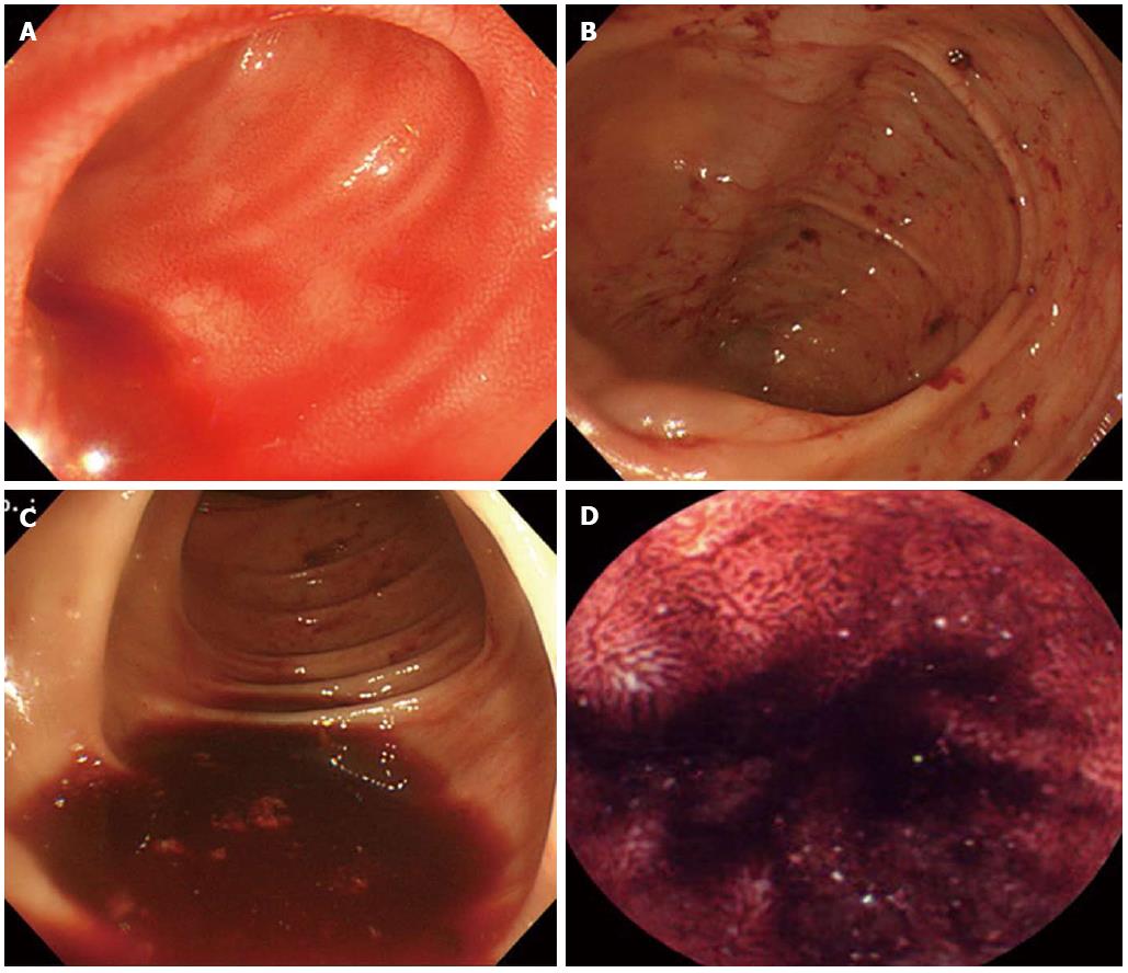
Figure 1 Colonoscopy and video capsule endoscopy findings.
A-C: Colonoscopy shows fresh blood material that gushed from the small bowel; D: Subsequent video capsule endoscopy shows evidence of active and recent bleeding in the ileum.
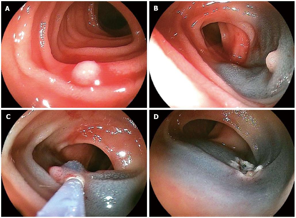
Figure 2 Double balloon enteroscopy findings.
A: Double balloon enteroscopy shows a small, whitish polypoid lesion with active bleeding in the distal ileum; B, C: After submucosal injection of a saline-epinephrine mixture, polypectomy was performed; D: After the procedure, argon plasma coagulation was performed on the post-polypectomy ulcer to achieve hemostasis.
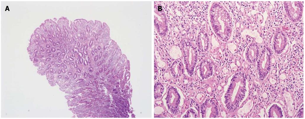
Figure 3 Histopathologic findings of intestinal lymphangiectasia.
A, B: Microscopic examination shows dilated lymphatic channels in the lamina propria (hematoxylin and eosin, × 40). Protein-rich fluid can escape from these channels into the extracellular space of the lamina propria and ultimately into the gut lumen (hematoxylin and eosin, × 200).
- Citation: Park MS, Lee BJ, Gu DH, Pyo JH, Kim KJ, Lee YH, Joo MK, Park JJ, Kim JS, Bak YT. Ileal polypoid lymphangiectasia bleeding diagnosed and treated by double balloon enteroscopy. World J Gastroenterol 2013; 19(45): 8440-8444
- URL: https://www.wjgnet.com/1007-9327/full/v19/i45/8440.htm
- DOI: https://dx.doi.org/10.3748/wjg.v19.i45.8440


