Copyright
©2013 Baishideng Publishing Group Co.
World J Gastroenterol. Dec 7, 2013; 19(45): 8342-8348
Published online Dec 7, 2013. doi: 10.3748/wjg.v19.i45.8342
Published online Dec 7, 2013. doi: 10.3748/wjg.v19.i45.8342
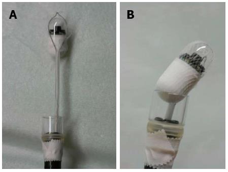
Figure 1 A capsule endoscope in a net device.
A: A capsule endoscope in a net device for foreign body extraction; B: A capsule endoscope in a net device for foreign body extraction; the device has been retracted and is seated firmly against a clear plastic hood attached to the tip of the upper gastrointestinal endoscope.
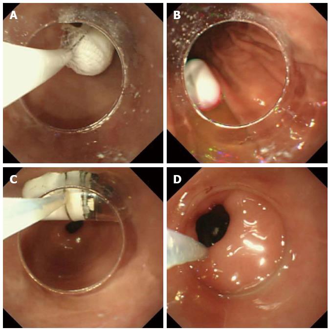
Figure 2 Capsule endoscope.
A: The capsule endoscope was delivered into the stomach with endoscopic assistance; B: The capsule endoscope was released into the stomach; C: The capsule endoscope was caught by the polypectomy snare; D: The capsule endoscope was introduced into the duodenal bulb with the snare and was then pushed with the plastic hood into the proximal duodenum.
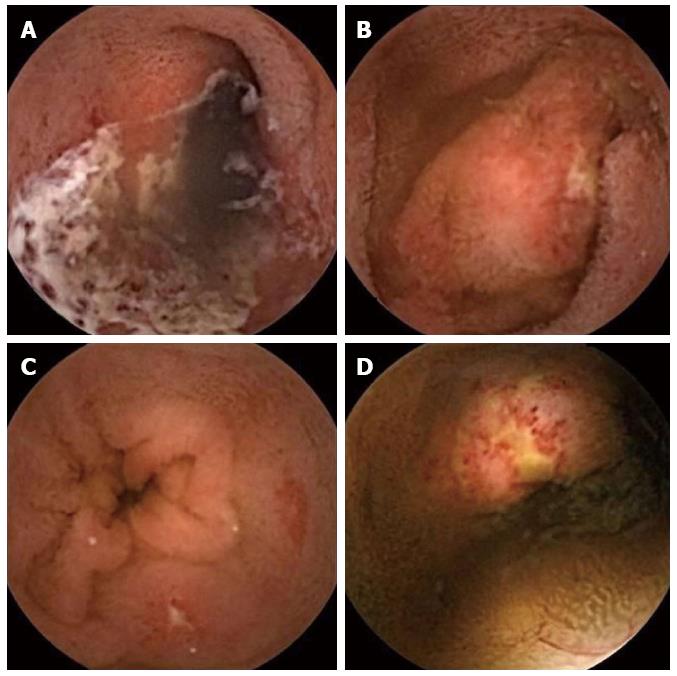
Figure 3 Three-year-old girl with Henoch-Schonlein purpura.
A: Extended ulcer; B, C and D: Multiple erosions and ulcers.
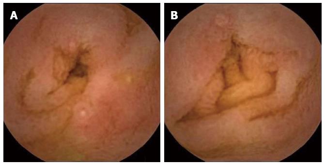
Figure 4 Ten-month-old girl with allergic gastroenteropathy (A and B) caused by the ingestion of cow’s milk.
- Citation: Oikawa-Kawamoto M, Sogo T, Yamaguchi T, Tsunoda T, Kondo T, Komatsu H, Inui A, Fujisawa T. Safety and utility of capsule endoscopy for infants and young children. World J Gastroenterol 2013; 19(45): 8342-8348
- URL: https://www.wjgnet.com/1007-9327/full/v19/i45/8342.htm
- DOI: https://dx.doi.org/10.3748/wjg.v19.i45.8342


