Copyright
©2013 Baishideng Publishing Group Co.
World J Gastroenterol. Nov 21, 2013; 19(43): 7813-7815
Published online Nov 21, 2013. doi: 10.3748/wjg.v19.i43.7813
Published online Nov 21, 2013. doi: 10.3748/wjg.v19.i43.7813
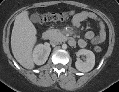
Figure 1 Contrast-enhanced computed tomography scan.
It shows decreased attenuation within the superior mesenteric vein (arrow), immediately below the portal confluence, compatible with venous thrombosis.
- Citation: Karmacharya P, Aryal MR, Donato A. Mesenteric vein thrombosis in a patient heterozygous for factor V Leiden and G20210A prothrombin genotypes. World J Gastroenterol 2013; 19(43): 7813-7815
- URL: https://www.wjgnet.com/1007-9327/full/v19/i43/7813.htm
- DOI: https://dx.doi.org/10.3748/wjg.v19.i43.7813


