Copyright
©2013 Baishideng Publishing Group Co.
World J Gastroenterol. Nov 21, 2013; 19(43): 7586-7593
Published online Nov 21, 2013. doi: 10.3748/wjg.v19.i43.7586
Published online Nov 21, 2013. doi: 10.3748/wjg.v19.i43.7586
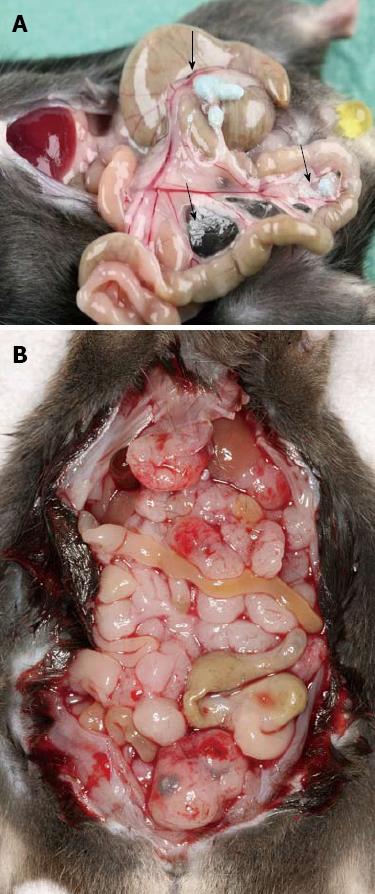
Figure 1 Peritoneal metastasis.
A: Beads accumulate in the mesentery of the small bowel (arrors). Animals show a complete tumor remission; B: Control animal with disseminated peritoneal carcinomatosis induced by EGFP-C-26 cells.
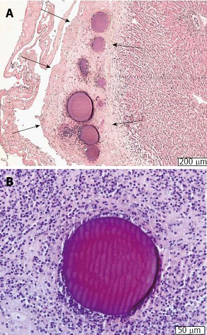
Figure 2 Doxorubicin-eluting beads.
A: HE-stained, magnification × 50: A layer of fibrin (between arrows) immobilizing the doxorubicin-loaded DC beads (DOXDEB™) on the livers surface; B: HE-stained, magnification × 100: DOXDEB™ in layer of fibrin and surrounded by lymphocytes immobilizing it on mesenteric connective tissue.
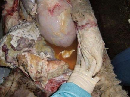
Figure 3 Chemical peritonitis.
Autopsy of an animal 28 d after installation of ip doxorubicin-loaded drug-eluting beads: Situs with greater omentum and intestinal loops with fibrinous adhesions, amber-colored ascitic fluid.
- Citation: Binder S, Lewis AL, Löhr JM, Keese M. Extravascular use of drug-eluting beads: A promising approach in compartment-based tumor therapy. World J Gastroenterol 2013; 19(43): 7586-7593
- URL: https://www.wjgnet.com/1007-9327/full/v19/i43/7586.htm
- DOI: https://dx.doi.org/10.3748/wjg.v19.i43.7586


