Copyright
©2013 Baishideng Publishing Group Co.
World J Gastroenterol. Nov 14, 2013; 19(42): 7472-7475
Published online Nov 14, 2013. doi: 10.3748/wjg.v19.i42.7472
Published online Nov 14, 2013. doi: 10.3748/wjg.v19.i42.7472
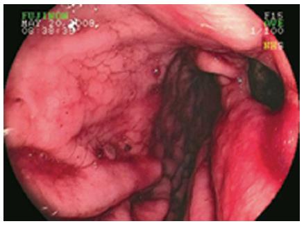
Figure 1 Esophagogastroduodenoscopy image showing the fistulous opening of the hepatocellular carcinoma on the lesser curvature of the stomach.
Figure 2 Computerized tomography images of the abdomen.
A: Showing large mass in the left lobe of the liver and oral contrast traced into liver (arrow) through fistulous opening in stomach; B: Showing large mass in the left lobe of the liver and fistulous communication between the liver mass and the stomach; C: Showing large mass in the left lobe of the liver and fistulous communication between the liver mass and the stomach. A: Anterior R: Right; L: Left; P: Posterior.
- Citation: Sayana H, Yousef O, Clarkston WK. Massive upper gastrointestinal hemorrhage due to invasive hepatocellular carcinoma and hepato-gastric fistula. World J Gastroenterol 2013; 19(42): 7472-7475
- URL: https://www.wjgnet.com/1007-9327/full/v19/i42/7472.htm
- DOI: https://dx.doi.org/10.3748/wjg.v19.i42.7472


