Copyright
©2013 Baishideng Publishing Group Co.
World J Gastroenterol. Nov 14, 2013; 19(42): 7241-7246
Published online Nov 14, 2013. doi: 10.3748/wjg.v19.i42.7241
Published online Nov 14, 2013. doi: 10.3748/wjg.v19.i42.7241
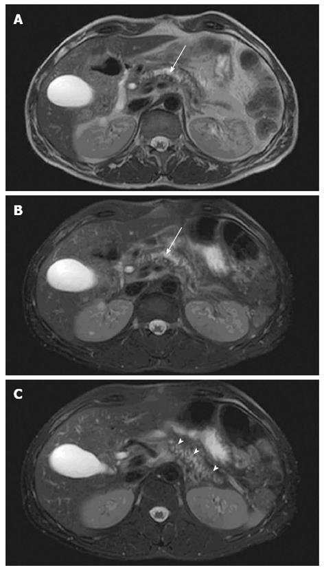
Figure 1 Pancreatic morphology.
Axial T2-weighted magnetic resonance imaging views showing glandular atrophy (A), dilated irregular duct (B, arrow) and irregular side-branches (C, arrow heads) in a patient with chronic pancreatitis.
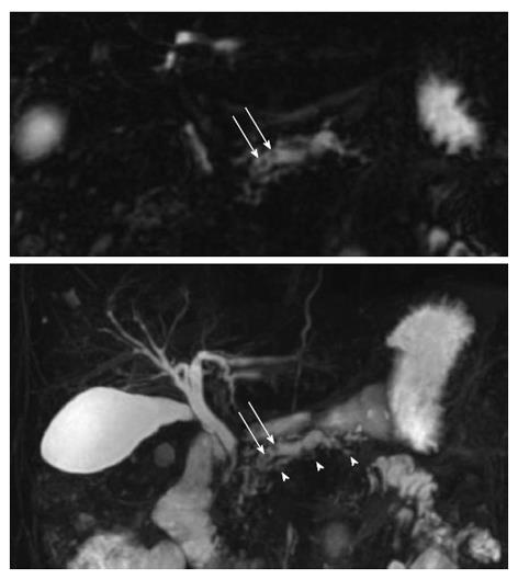
Figure 2 Magnetic resonance cholangiopancreatography.
Upper figure displays a single coronal image and the lower figure a 3D view of the pancreato-biliary tree. In this chronic pancreatitis patient the dilated irregular main duct has irregular side branches (arrow heads) and contains multiple rounded filling defects (arrows).
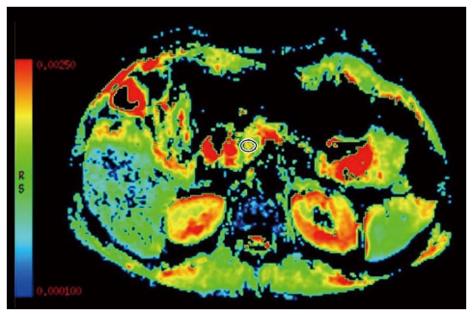
Figure 3 Diffusion weighted imaging.
The apparent diffusion coefficient map of a chronic pancreatitis patient is shown with a measuring region of interest positioned in the pancreatic head to assess the degree of parenchymal fibrosis (red is high and blue low water diffusion).
- Citation: Hansen TM, Nilsson M, Gram M, Frøkjær JB. Morphological and functional evaluation of chronic pancreatitis with magnetic resonance imaging. World J Gastroenterol 2013; 19(42): 7241-7246
- URL: https://www.wjgnet.com/1007-9327/full/v19/i42/7241.htm
- DOI: https://dx.doi.org/10.3748/wjg.v19.i42.7241


