Copyright
©2013 Baishideng Publishing Group Co.
World J Gastroenterol. Nov 7, 2013; 19(41): 7160-7167
Published online Nov 7, 2013. doi: 10.3748/wjg.v19.i41.7160
Published online Nov 7, 2013. doi: 10.3748/wjg.v19.i41.7160
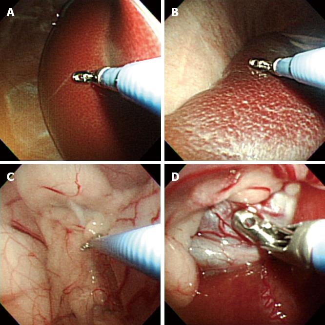
Figure 1 Peritonescopy and endoscopic biopsy of the intraperitoneal organs.
A: Liver; B: Spleen; C: Abdominal wall; D: Omentum.
Figure 2 Forward-viewing endoscopic ultrasound image of the intraperitoneal organs and endoscopic ultrasound-guided fine needle aspiration using a 19G aspiration needle (white arrow).
A: Liver; B: Kidney; C: Spleen.
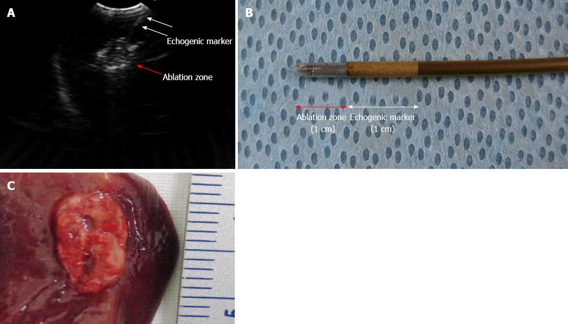
Figure 3 Endoscopic ultrasound-guided radio frequency ablation using an 18G radiofrequency ablation needle on the hepatic parenchyma.
A: Radiofrequency ablation (RFA) needle (white arrow) in the hepatic parenchyma with echogenic ablation zone (red arrow); B: RFA needle tip with ablation zone (red arrow) echogenic marker (white arrow); C: Gross pathology of ablated tissue in the liver parenchyma.
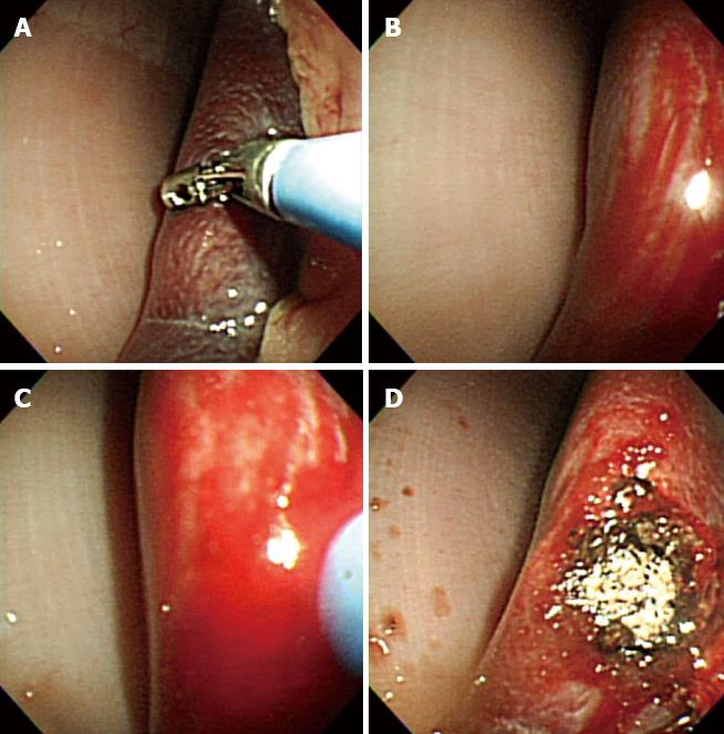
Figure 4 Argon plasma coagulation for hemostatic control of artificially induced bleeding at the spleen.
A: Bleeding induced by a biopsy forcep; B: Surface bleeding seen at the spleen; C: Argon plasma coagulation catheter introduced to achieve thermal coagulation; D: Hemostasis successfully achieved.
- Citation: Jeong SU, Aizan H, Song TJ, Seo DW, Kim SH, Park DH, Lee SS, Lee SK, Kim MH. Forward-viewing endoscopic ultrasound-guided NOTES interventions: A study on peritoneoscopic potential. World J Gastroenterol 2013; 19(41): 7160-7167
- URL: https://www.wjgnet.com/1007-9327/full/v19/i41/7160.htm
- DOI: https://dx.doi.org/10.3748/wjg.v19.i41.7160


