Copyright
©2013 Baishideng Publishing Group Co.
World J Gastroenterol. Nov 7, 2013; 19(41): 7089-7096
Published online Nov 7, 2013. doi: 10.3748/wjg.v19.i41.7089
Published online Nov 7, 2013. doi: 10.3748/wjg.v19.i41.7089
Figure 1 Screening endoscopy.
A-C: Endoscopically suspected short-segment Barrett’s esophagus (< 3 cm).
Figure 2 Magnifying endoscopy.
A: Magnifying endoscopy up to × 80 (Olympus GIF-Q240Z, × 80); B: Magnified image of the short-segment Barrett’s esophagus.
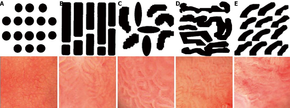
Figure 3 Classification of pit-pattern of Barrett’s esophagus by Magnifying endoscopy (Endo’s classification).
A:I (small round); B: II (straight); C: III (long oval); D: IV (tubular); E: V (villous).
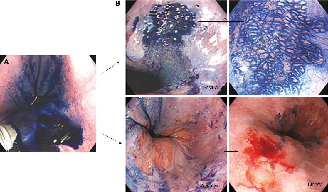
Figure 4 Methylene blue chromoendoscopy.
A: 0.5% solution of methylene blue (MB) was sprayed over the columnar mucosa; B: Biopsies were obtained from the regions previously observed by magnifying endoscopy and MB chromoendoscopy.
Figure 5 Histological diagnosis.
A: Fundic type (HE stain, × 200); B: Cardiac type (HE stain, × 200); C: Specialized intestinal metaplasia (HE stain, × 400).
Figure 6 Distributions of pit-pattern, methylene blue staining, histologic diagnosis.
A: Pit-pattern; B: Methylene blue staining; C: Histologic diagnosis. SIM: Specialized intestinal metaplasia.
- Citation: Ham NS, Jang JY, Ryu SW, Kim JH, Park EJ, Lee WC, Shim KY, Jeong SW, Kim HG, Lee TH, Jeon SR, Cho JH, Cho JY, Jin SY, Lee JS. Magnifying endoscopy for the diagnosis of specialized intestinal metaplasia in short-segment Barrett’s esophagus. World J Gastroenterol 2013; 19(41): 7089-7096
- URL: https://www.wjgnet.com/1007-9327/full/v19/i41/7089.htm
- DOI: https://dx.doi.org/10.3748/wjg.v19.i41.7089


