Copyright
©2013 Baishideng Publishing Group Co.
World J Gastroenterol. Jan 28, 2013; 19(4): 542-549
Published online Jan 28, 2013. doi: 10.3748/wjg.v19.i4.542
Published online Jan 28, 2013. doi: 10.3748/wjg.v19.i4.542
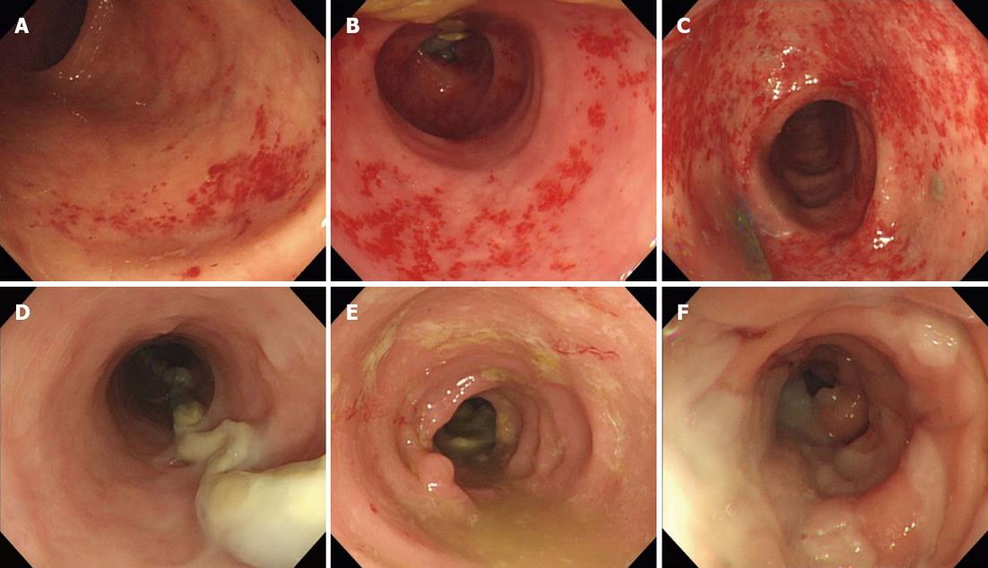
Figure 1 Example of major endoscopic findings in diversion colitis according to relative severity.
A-C: Mucosal hemorrhage (A: Score 1; B: Score 2; C: Score3); D-F: Edema (D: Score 1; E: Score 2; F: Score 3).
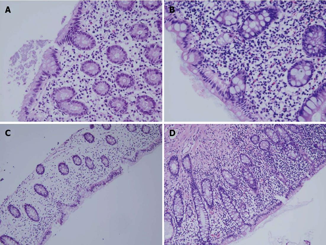
Figure 2 Example of major histological findings in diversion colitis according to relative severity.
A, B: Eosinophilic infiltration including numerous cytoplasmic granules and bilobed nucleus (A: Score 1; B: Score 2; hematoxylin and eosin stain × 400); C, D: Chronic inflammation with mononuclear cell such as macrophage,lymaphocytes and plasma cells (C: Score 1; D: Score 2; hematoxylin and eosin stain × 200).
- Citation: Son DN, Choi DJ, Woo SU, Kim J, Keom BR, Kim CH, Baek SJ, Kim SH. Relationship between diversion colitis and quality of life in rectal cancer. World J Gastroenterol 2013; 19(4): 542-549
- URL: https://www.wjgnet.com/1007-9327/full/v19/i4/542.htm
- DOI: https://dx.doi.org/10.3748/wjg.v19.i4.542


