Copyright
©2013 Baishideng Publishing Group Co.
World J Gastroenterol. Jan 28, 2013; 19(4): 536-541
Published online Jan 28, 2013. doi: 10.3748/wjg.v19.i4.536
Published online Jan 28, 2013. doi: 10.3748/wjg.v19.i4.536
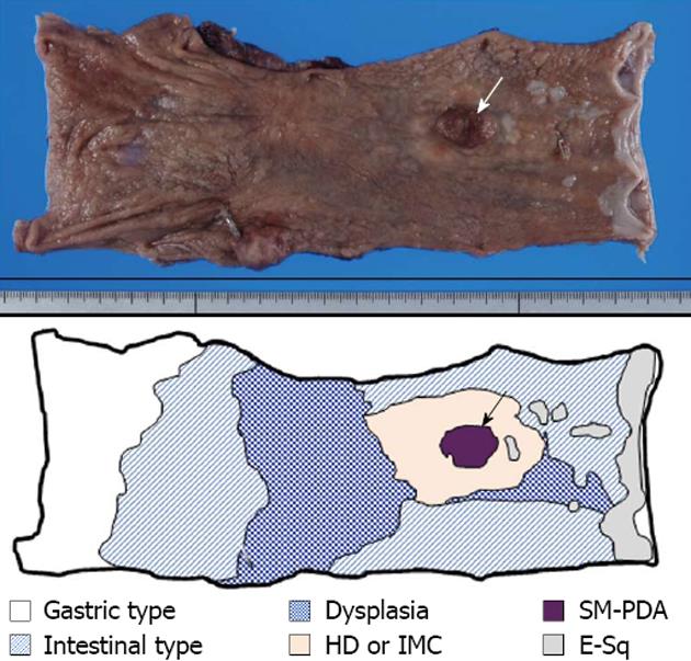
Figure 1 Macroscopic findings of excised esophagus.
Surgical specimen shows the presence of superficial spreading carcinoma, extending between 30 mm from the oral and 105 mm from the anal surgical margins. The superficial spreading region is mostly composed of high-grade dysplasia (HD), or intramucosal adenocarcinoma (IMC), surrounded by dysplastic change (E-sq low-grade dysplasia). Inside this superficial spreading region, one observable elevated nodule (arrow) is composed of solid and submucosal invasive poorly-differentiated invasive adenocarcinoma (SM-PDA) with lymphatic invasion. The background non-neoplastic esophageal mucosa is extensively replaced by glandular mucosa with and without intestinal metaplasia.
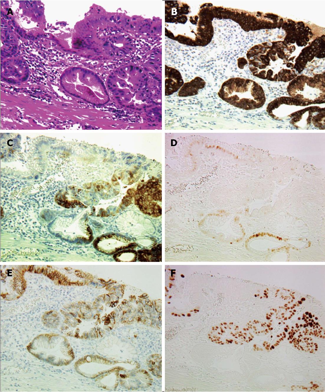
Figure 2 Histological findings of transitional area between intestinal metaplasia and high-grade dysplasia or intramucosal adenocarcinoma (× 200).
A: Hematoxylin and eosin staining of transitional area. Intestinal metaplasia (IM) stretches from the upper left to the lower right corner; B: Mucin (MUC) 5AC immunostaining. Strong MUC5AC expression is observed in both IM and high-grade dysplasia (HD), or intramucosal adenocarcinoma (IMC) areas; C: MUC6 immunostaining. MUC6 expression is observed mostly in parts of the HD or IMC areas; D: Caudal type homebox transcription factor 2 (Cdx2) immunostaining. Cdx2 expression is observed only in the nuclei of the cells in the IM area; E: E-cad immunostaining. E-cad expression is observed on the membranes of cells in both IM and HD or IMC areas; F: p53 immunostaining. Strong p53 expression is observed in the nuclei of the cells in the HD or IMC area.
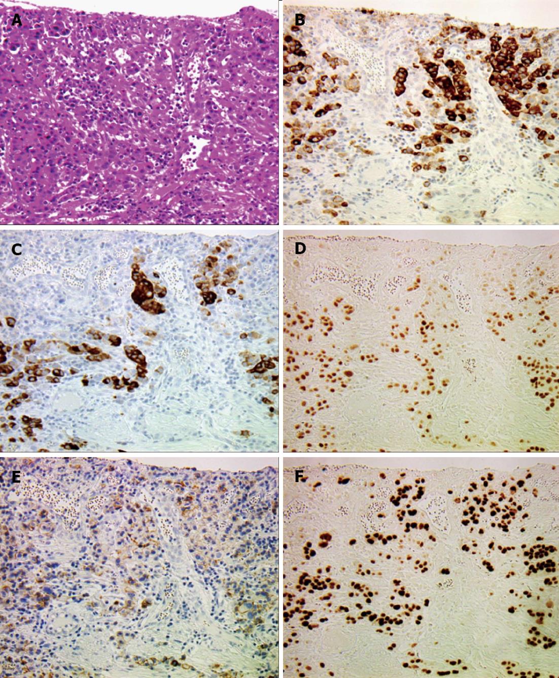
Figure 3 Histological findings of diffuse poorly-differentiated invasive adenocarcinoma (× 200).
A: Hematoxylin and eosin staining of poorly-differentiated invasive adenocarcinoma (PDA). Cancer cells are diffusely scattered with prominent stromal reaction; B: Mucin (MUC) 5AC; C: MUC6; D: Caudal type homebox transcription factor 2 expression is positive in the PDA area; E: E-cad expression is markedly reduced in the PDA area; F: Strong p53 expression is observed in the nuclei of the cells in the PDA area.
Figure 4 Detection of methylated cytosine by methylation-specific polymerase chain reaction analysis of caudal type homebox transcription factor 2 CpG-island region.
Tissue samples were stained with E-cadherin and caudal type homebox transcription factor 2 (Cdx2) to assist cell identification. Cells, either isolated from intestinal metaplasia, intramucosal adenocarcinoma (IMC) or submucosal invasive poorly-differentiated adenocarcinoma (SM-PDA) by laser-assisted microdissection, were subjected to bisulfite conversion and subsequent methylation-specific polymerase chain reaction (MSP). MSP products using primers that specifically amplify only unmethylated DNA are indicated by visible polymerase chain reaction products in line unmethylated pattern (U), while visible polymerase chain reaction products in line methylated pattern (M) indicate those amplified by primers specific for methylated DNA. In intestinal-type metaplasia sections, MSP shows a U pattern, while MSP shows U patterns with partial M patterns in high-grade dysplasia or IMC sections. MSP shows a mostly M pattern in PDA where strong Cdx2 expression is observed (Figure 3D).
- Citation: Makita K, Kitazawa R, Semba S, Fujiishi K, Nakagawa M, Haraguchi R, Kitazawa S. Cdx2 expression and its promoter methylation during metaplasia-dysplasia-carcinoma sequence in Barrett's esophagus. World J Gastroenterol 2013; 19(4): 536-541
- URL: https://www.wjgnet.com/1007-9327/full/v19/i4/536.htm
- DOI: https://dx.doi.org/10.3748/wjg.v19.i4.536


