Copyright
©2013 Baishideng Publishing Group Co.
World J Gastroenterol. Oct 14, 2013; 19(38): 6500-6504
Published online Oct 14, 2013. doi: 10.3748/wjg.v19.i38.6500
Published online Oct 14, 2013. doi: 10.3748/wjg.v19.i38.6500
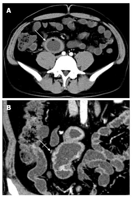
Figure 1 Computed tomography enterography scan.
A: Transverse view showed suspicious ileal intussusception (white arrow) in the right lower quadrant; B: Coronal view revealed a similar result (white arrow).
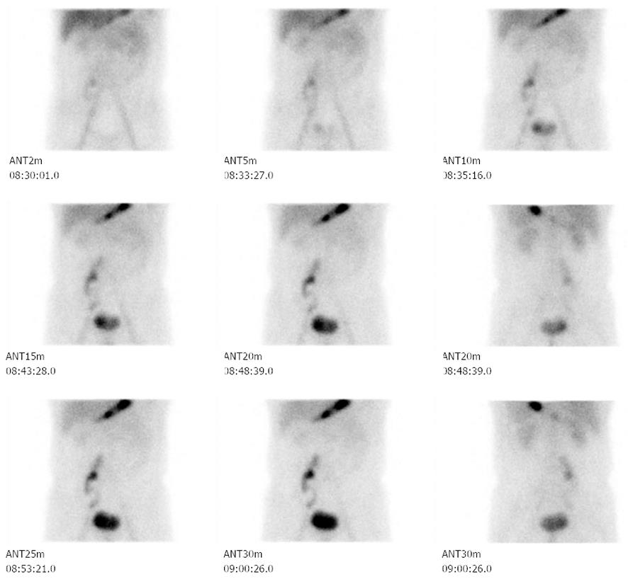
Figure 2 Tc-99m pertechnetate scintigraphy.
A cluster of stripy abnormal radio-activity was located in the right lower quadrant.
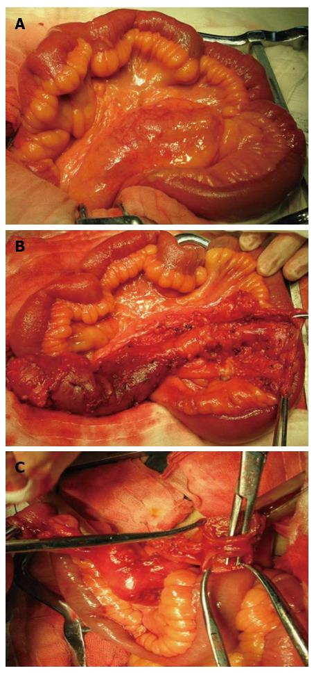
Figure 3 Exploratory laparotomy findings.
A: A 25-cm duplicating, tubular small intestinal segment was found arising from the ileal mesenteric margin; B: This duplication cyst was intimately attached to the native ileal segment located 15 cm proximal to ileocecal valve; C: this cyst had a blind end proximally and a completely patent orifice into the native ileal lumen distally.
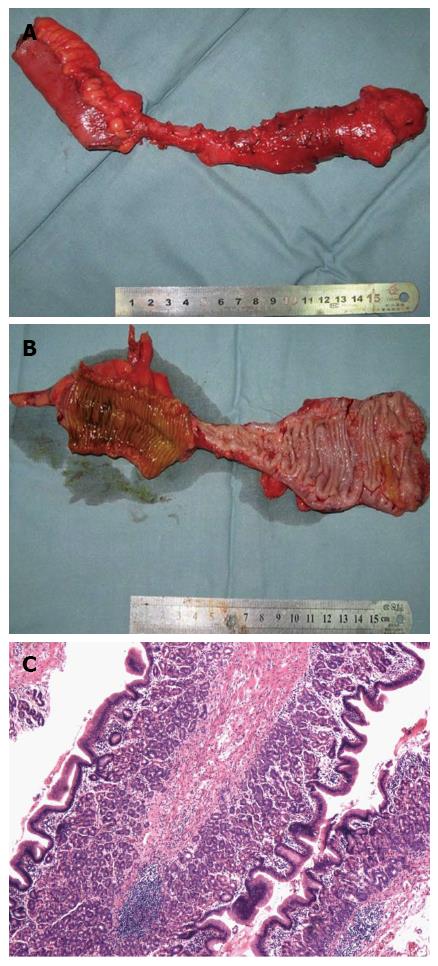
Figure 4 Gross and histological pathology of the resection specimen.
A: The resection specimen showed no signs of inflammation, infection, ulceration, hemorrhage, obstruction, or malignant transformation; B: The mucosal layer of the duplication cyst was lined with both small intestinal and gastric mucosae; C: Histology revealed that the duplication cyst was lined with ileal mucus glands and heterotopic gastric mucosae (hematoxylin-eosin, × 100).
- Citation: Li BL, Huang X, Zheng CJ, Zhou JL, Zhao YP. Ileal duplication mimicking intestinal intussusception: A congenital condition rarely reported in adult. World J Gastroenterol 2013; 19(38): 6500-6504
- URL: https://www.wjgnet.com/1007-9327/full/v19/i38/6500.htm
- DOI: https://dx.doi.org/10.3748/wjg.v19.i38.6500


