Copyright
©2013 Baishideng Publishing Group Co.
World J Gastroenterol. Oct 14, 2013; 19(38): 6329-6347
Published online Oct 14, 2013. doi: 10.3748/wjg.v19.i38.6329
Published online Oct 14, 2013. doi: 10.3748/wjg.v19.i38.6329
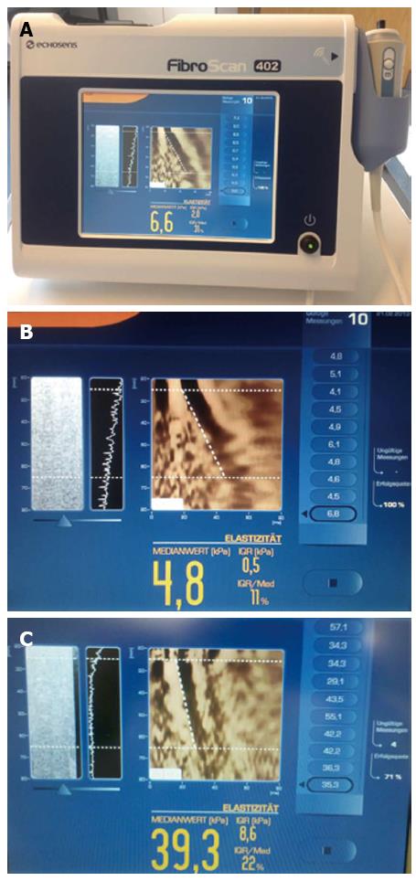
Figure 1 Transient elastography (Fibroscan®, A) for the evaluation of normal liver (B) and liver cirrhosis (C).
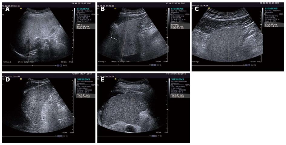
Figure 2 Point-shear wave elastography with acoustic radiation force impulse for the evaluation of histologically proved liver fibrosis and cirrhosis.
A: F0; B: F1; C: F2; D: F3; E: F4 = cirrhosis.
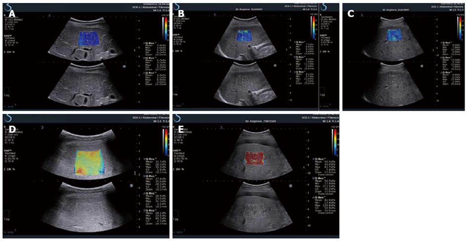
Figure 3 2D-shear wave elastography with supersonic shear imaging for the evaluation of histologically proved liver fibrosis and cirrhosis.
A: F0; B: F1; C: F2; D: F3; E: F4 = cirrhosis.
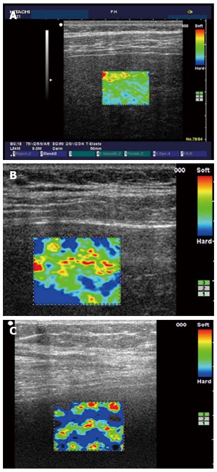
Figure 4 Strain elastography for the evaluation of histologically proved liver fibrosis and cirrhosis.
A: F0; B: F2; C: F4 = cirrhosis.
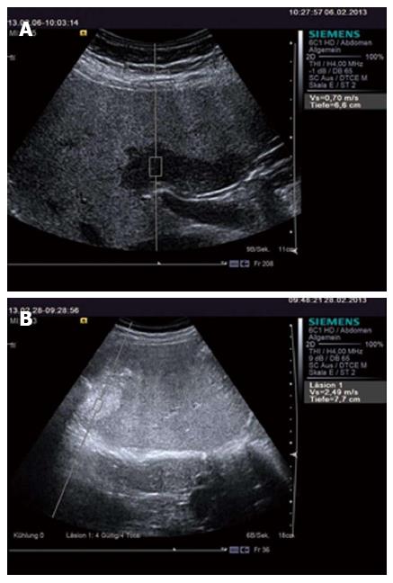
Figure 5 Point-shear wave elastography with acoustic radiation force impulse for evaluation of focal liver fatty lesion (A) and liver metastasis (B).
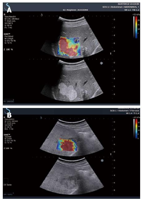
Figure 6 2D shear wave elastography with supersonic shear imaging for the evaluation of liver hemangioma (A) and metastasis (B).
- Citation: Cui XW, Friedrich-Rust M, Molo CD, Ignee A, Schreiber-Dietrich D, Dietrich CF. Liver elastography, comments on EFSUMB elastography guidelines 2013. World J Gastroenterol 2013; 19(38): 6329-6347
- URL: https://www.wjgnet.com/1007-9327/full/v19/i38/6329.htm
- DOI: https://dx.doi.org/10.3748/wjg.v19.i38.6329


