Copyright
©2013 Baishideng Publishing Group Co.
World J Gastroenterol. Oct 7, 2013; 19(37): 6292-6295
Published online Oct 7, 2013. doi: 10.3748/wjg.v19.i37.6292
Published online Oct 7, 2013. doi: 10.3748/wjg.v19.i37.6292
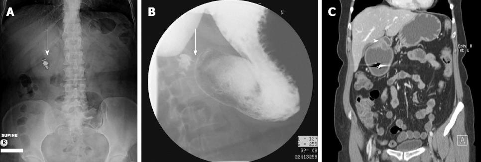
Figure 1 Radiologic imaging.
A: Retained video capsule on abdominal roentogram (arrow); B: Barium upper gastrointestinal series (arrow) shows pronounced delay in gastric emptying with a narrowed slightly elongated pylorus and a distended gastric antrum; C: Computed enterography of the abdomen shows retained video capsule, distended antrum of the stomach, and abnormally dense, eccentric, and significantly narrowed pylorus (arrow).
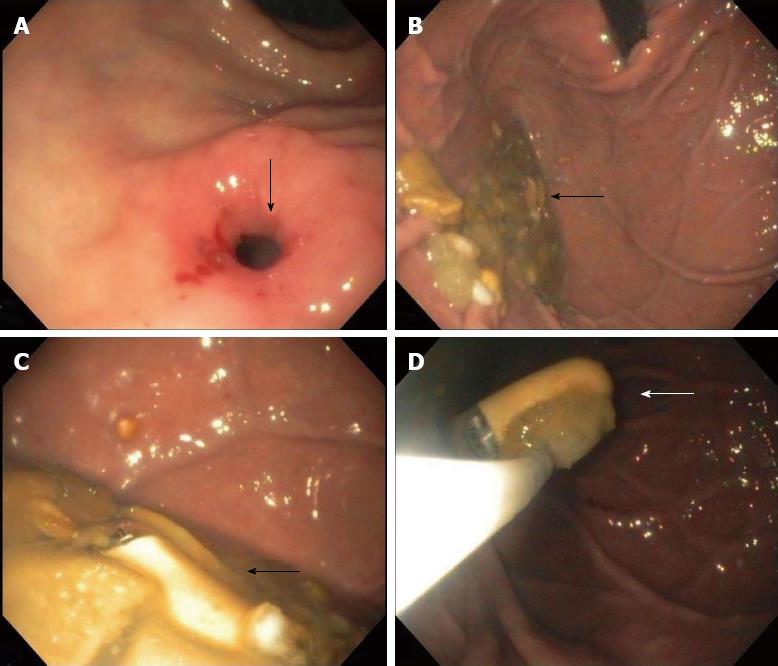
Figure 2 Endoscopic findings.
A: Eccentric hypertrophic pyloric stenosis (arrow); B: Retroflexed view of the gastric fundus shows freely floating 3 years old video capsule in pool of retained semi-digested food particles (arrow); C: Close up view of retained capsule (arrow); D: Endoscopic retrieval with Roth Net (arrow).
- Citation: Gurvits GE, Tan A, Volkov D. Video capsule endoscopy and CT enterography in diagnosing adult hypertrophic pyloric stenosis. World J Gastroenterol 2013; 19(37): 6292-6295
- URL: https://www.wjgnet.com/1007-9327/full/v19/i37/6292.htm
- DOI: https://dx.doi.org/10.3748/wjg.v19.i37.6292


