Copyright
©2013 Baishideng Publishing Group Co.
World J Gastroenterol. Sep 21, 2013; 19(35): 5925-5928
Published online Sep 21, 2013. doi: 10.3748/wjg.v19.i35.5925
Published online Sep 21, 2013. doi: 10.3748/wjg.v19.i35.5925
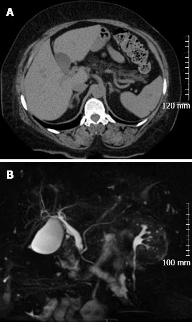
Figure 1 Radiological images of the abdomen.
A: Computed tomography scan without contrast demonstrating the gallbladder to the left of the falciform ligament; B: Magnetic resonance cholangiopancreatography showing a dilated common bile duct on coronal view.
- Citation: Iskandar ME, Radzio A, Krikhely M, Leitman IM. Laparoscopic cholecystectomy for a left-sided gallbladder. World J Gastroenterol 2013; 19(35): 5925-5928
- URL: https://www.wjgnet.com/1007-9327/full/v19/i35/5925.htm
- DOI: https://dx.doi.org/10.3748/wjg.v19.i35.5925


