Copyright
©2013 Baishideng Publishing Group Co.
World J Gastroenterol. Aug 28, 2013; 19(32): 5377-5380
Published online Aug 28, 2013. doi: 10.3748/wjg.v19.i32.5377
Published online Aug 28, 2013. doi: 10.3748/wjg.v19.i32.5377
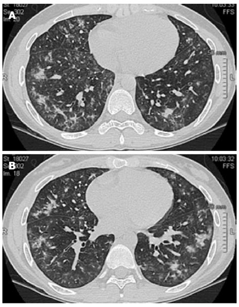
Figure 1 High-resolution computed tomography at hospital admission in the Pneumology Unit.
High-resolution computed tomography of the thorax revealed bilateral shadowing nodules and adjacent interstitial (A) thickening with a predominant distribution in the middle and basal regions and relative sparing of the apices (B). R: Right; L: Left.
- Citation: Caccaro R, Savarino E, D’Incà R, Sturniolo GC. Noninfectious interstitial lung disease during infliximab therapy: Case report and literature review. World J Gastroenterol 2013; 19(32): 5377-5380
- URL: https://www.wjgnet.com/1007-9327/full/v19/i32/5377.htm
- DOI: https://dx.doi.org/10.3748/wjg.v19.i32.5377


