Copyright
©2013 Baishideng Publishing Group Co.
World J Gastroenterol. Jan 21, 2013; 19(3): 422-425
Published online Jan 21, 2013. doi: 10.3748/wjg.v19.i3.422
Published online Jan 21, 2013. doi: 10.3748/wjg.v19.i3.422
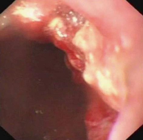
Figure 1 Gastroscopic examination revealed a large irregular ulcer with apophysis and erosive hemorrhage located on the posterior wall of fundus and lesser curvature.
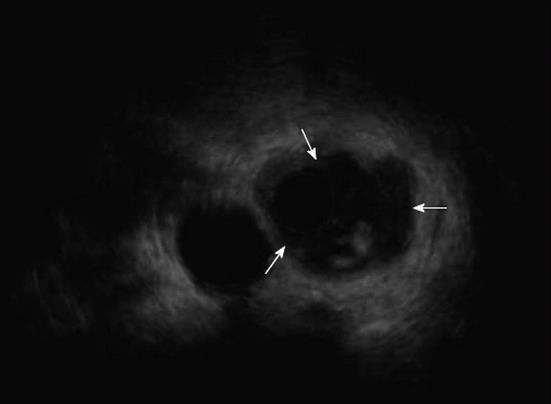
Figure 2 Endoscopic ultrasonography showed thickening about the semi-perimeter of the gastric fundal mucosa and a heterogenous hypoechoic mass, the whole layer of the local wall involved (arrows).
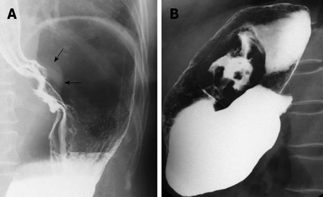
Figure 3 Double contrast examination demonstrated the shadow of soft tissue mass around the cardia and lesser curvature of stomach (arrows) (A), and filling defect, intracavitary niches, destruction of mucosa, as well as rigidity of the local wall were seen (B).
The abdominal part of esophagus was involved.
Figure 4 Abdominal computed tomography scan showed focal irregular thickened gastric wall and a ulcerated soft tissue mass formation with heterogeneously obvious enhancement in the lesser gastric curvature and fundus area.
A: Plain scan; B: Contrast-enhanced scan; C: Coronal multiplanar reconstruction of enhancement scan.
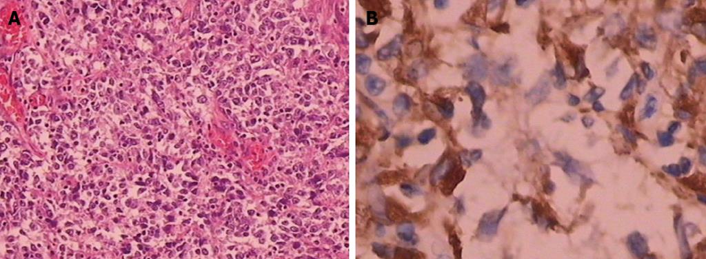
Figure 5 Histopathologic exam showed the tumor cells were marked inequality in the sizes, the cytoplasm was abundant and eosinophilic, the nuclei were large, round to oval, bizarre multinuclear giant cells were scattered.
The tumor cells expressed CD68. A: Hematoxylin and eosin ×100; B: Immunohistochemical staining for CD68 ×400.
- Citation: Shen XZ, Liu F, Ni RJ, Wang BY. Primary histiocytic sarcoma of the stomach: A case report with imaging findings. World J Gastroenterol 2013; 19(3): 422-425
- URL: https://www.wjgnet.com/1007-9327/full/v19/i3/422.htm
- DOI: https://dx.doi.org/10.3748/wjg.v19.i3.422


