Copyright
©2013 Baishideng Publishing Group Co.
World J Gastroenterol. Jan 21, 2013; 19(3): 339-346
Published online Jan 21, 2013. doi: 10.3748/wjg.v19.i3.339
Published online Jan 21, 2013. doi: 10.3748/wjg.v19.i3.339
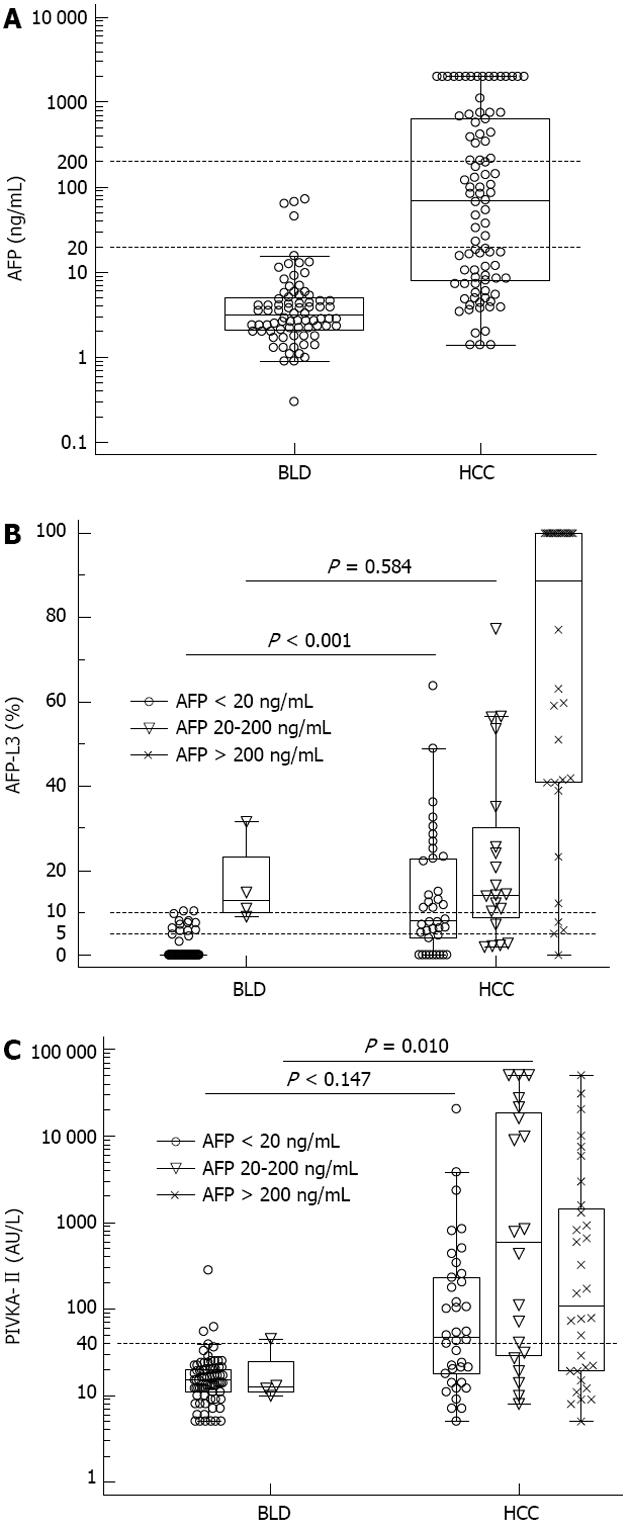
Figure 1 α-fetoprotein (A), α-fetoprotein-L3 (B) and prothrombin induced by vitamin K absence-II (C) values in benign liver disease and hepatocellular carcinoma patients.
α-fetoprotein (AFP)-L3 and prothrombin induced by vitamin K absence (PIVKA)-II levels are plotted by AFP concentrations (AFP < 20 ng/mL, AFP 20-200 ng/mL and AFP > 200 ng/mL). In patients with AFP < 20 ng/mL, AFP-L3 levels were higher in hepatocellular carcinoma (HCC) (P < 0.001) (B). In patients with AFP 20-200 ng/mL, PIVKA-II showed higher levels in HCC (P = 0.010) (C). The horizontal dotted lines are cut-off values used for performance analysis in this study. BLD: Benign liver disease.
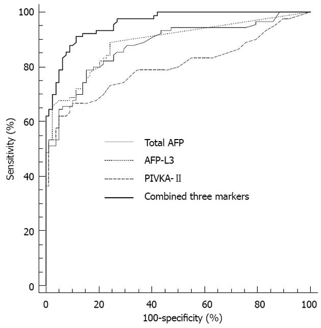
Figure 2 Receiver operating characteristic curves of total α-fetoprotein, α-fetoprotein-L3, prothrombin induced by vitamin K absence-II and three combined markers for the diagnosis of hepatocellular carcinoma in all patients.
The area under the curve values were 0.879 for total α-fetoprotein (AFP), 0.887 for AFP-L3, 0.801 for prothrombin induced by vitamin K absence (PIVKA)-II and 0.959 for the three combined markers.
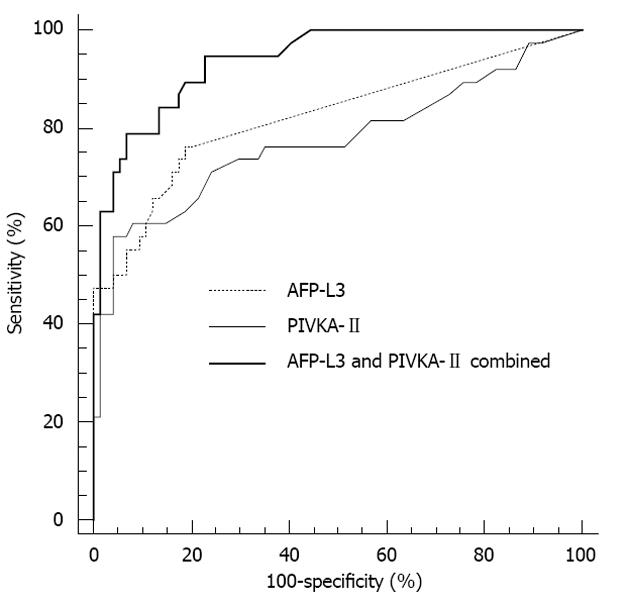
Figure 3 Receiver operating characteristic curves of α-fetoprotein-L3 and prothrombin induced by vitamin K absence-II for the diagnosis of hepatocellular carcinoma in patients with total α-fetoprotein < 20 ng/mL.
The area under the curve values were 0.824 for α-fetoprotein (AFP)-L3, 0.774 for prothrombin induced by vitamin K absence (PIVKA)-II and 0.939 for the two combined markers.
- Citation: Choi JY, Jung SW, Kim HY, Kim M, Kim Y, Kim DG, Oh EJ. Diagnostic value of AFP-L3 and PIVKA-II in hepatocellular carcinoma according to total-AFP. World J Gastroenterol 2013; 19(3): 339-346
- URL: https://www.wjgnet.com/1007-9327/full/v19/i3/339.htm
- DOI: https://dx.doi.org/10.3748/wjg.v19.i3.339


