Copyright
©2013 Baishideng Publishing Group Co.
World J Gastroenterol. Aug 7, 2013; 19(29): 4832-4835
Published online Aug 7, 2013. doi: 10.3748/wjg.v19.i29.4832
Published online Aug 7, 2013. doi: 10.3748/wjg.v19.i29.4832
Figure 1 Chest posterior to anterior of case 1.
The patient had undergone a right pneumonectomy.
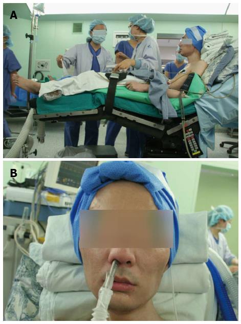
Figure 2 A 42-year-old male patient suffering from ankylosing spondylitis was admitted to our hospital presenting with right upper quadrant pain.
A: Operating position used for case 2; B: Awake fiberoptic intubation through the nasotracheal tree used for case 2.
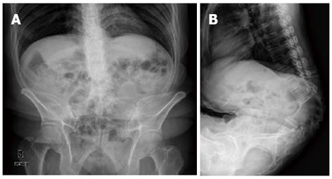
Figure 3 Thoracolumbar spine image for case 3.
The anteroposterior (A) and lateral (B) views are shown.
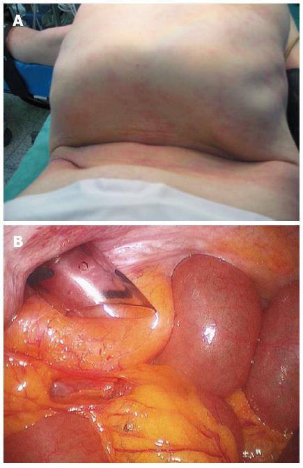
Figure 4 Insertion of the trocar in case 3.
A: The narrow abdomen provided a challenge for trocar insertion; B: Internal view of trocar placement in the narrow abdomen.
- Citation: Kim BS, Joo SH, Joh JH, Yi JW. Laparoscopic cholecystectomy in patients with anesthetic problems. World J Gastroenterol 2013; 19(29): 4832-4835
- URL: https://www.wjgnet.com/1007-9327/full/v19/i29/4832.htm
- DOI: https://dx.doi.org/10.3748/wjg.v19.i29.4832


