Copyright
©2013 Baishideng Publishing Group Co.
World J Gastroenterol. Aug 7, 2013; 19(29): 4651-4670
Published online Aug 7, 2013. doi: 10.3748/wjg.v19.i29.4651
Published online Aug 7, 2013. doi: 10.3748/wjg.v19.i29.4651
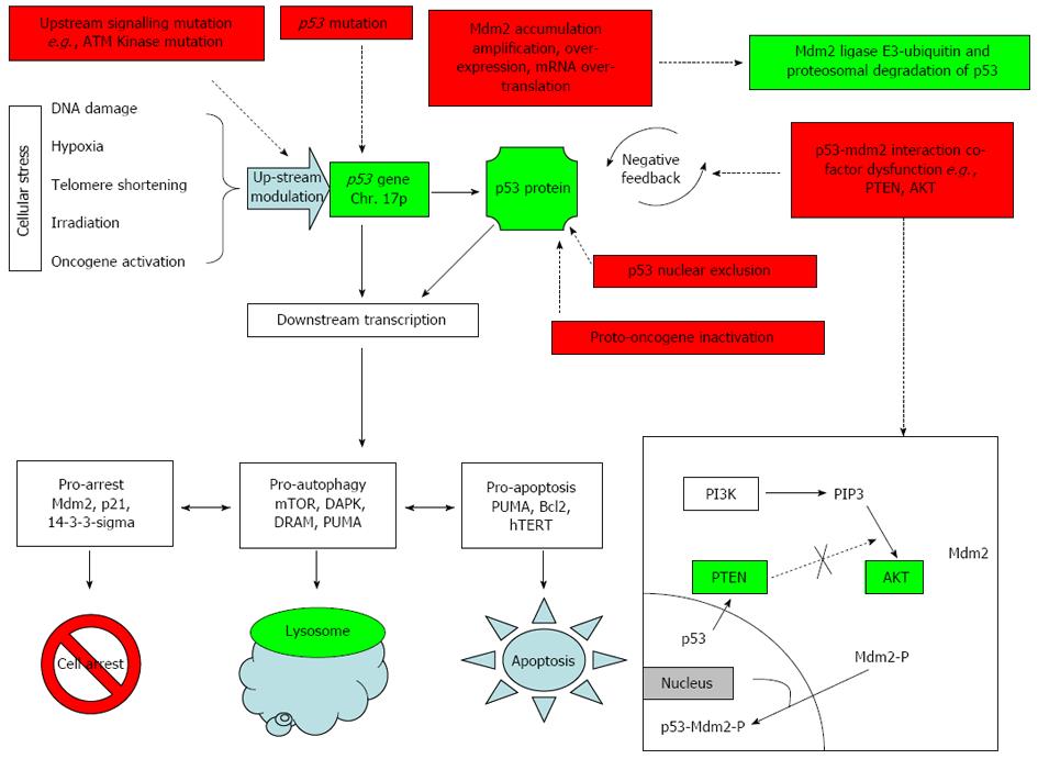
Figure 1 Normal p53 function highlighted in green boxes and blue figures as described.
Red boxes show mechanisms for inactivation; Normal p53 function highlighted in green and blue figures. PI3K: Phosphatidylinositol-3-kinase; PTEN: Phosphatase and tensin homolog; AKT: Protein kinase B; ATM: Ataxia telangiectasia mutated kinase; mTOR: Mammalian target of rapamycin; DAPK: Death-associated protein kinase; DRAM: Damage-regulated autophagy modulator; PUMA: p53-upregulated modulator of apoptosis; hTERT: Human telomerase reverse transcriptase.
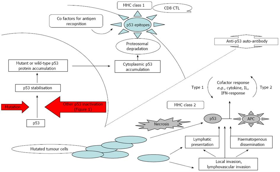
Figure 2 Proposed mechanisms of anti-p53 auto-antibody induction.
MHC: Major histocompatibility complex; CD8: Cluster of 8 differentiation; CTL: Cytotoxic T-cell; IL: Interleukin; APC: Antigen presenting cell.
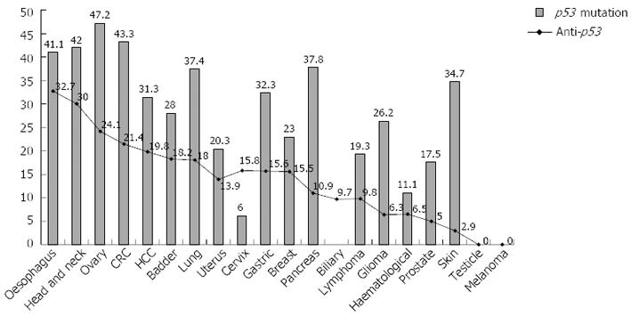
Figure 3 Percent p53 mutations (International Agency for Research on Cancer, 2008) and percent anti-p53 auto-antibody incidence calculated in this review.
r2 = 0.45, Correlation = 0.59. CRC: Colorectal cancer; HCC: Hepatocellular carcinoma.
-
Citation: Suppiah A, Greenman J. Clinical utility of anti-
p53 auto-antibody: Systematic review and focus on colorectal cancer. World J Gastroenterol 2013; 19(29): 4651-4670 - URL: https://www.wjgnet.com/1007-9327/full/v19/i29/4651.htm
- DOI: https://dx.doi.org/10.3748/wjg.v19.i29.4651


