Copyright
©2013 Baishideng Publishing Group Co.
World J Gastroenterol. Jul 21, 2013; 19(27): 4432-4436
Published online Jul 21, 2013. doi: 10.3748/wjg.v19.i27.4432
Published online Jul 21, 2013. doi: 10.3748/wjg.v19.i27.4432
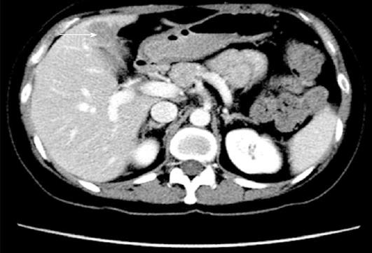
Figure 1 Surveillance computed tomography scan showing hypodense lesion in segment IVb of liver.
Cross-sectional image of the hypodense lesion in segment IVb of the liver (arrow).
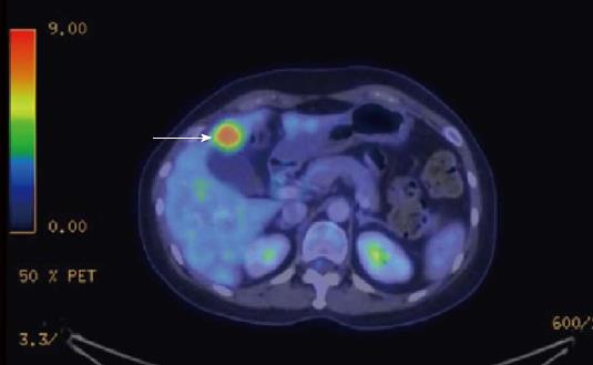
Figure 2 Computed tomography-positron emission tomography scan revealing the positron emission tomography avid lesion in segment IVb of the liver.
Cross-sectional image of the positron emission tomography avid lesion in segment IVb of the liver (standardized uptake value 7.9) (arrow).
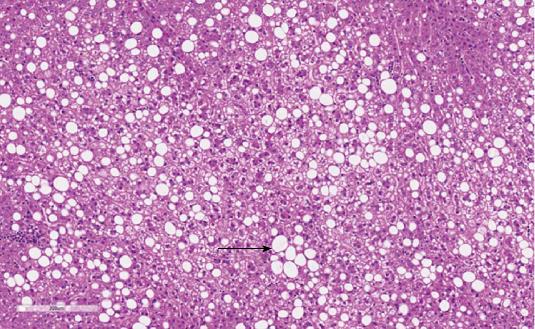
Figure 3 Representative histological slide of the hepatic adenoma.
Medium power HE of the hepatic adenoma shows trabeculae with normal thickness composed of hepatocytes with moderate to severe steatotic changes (arrow) and bland uniform nuclei. No large cell or small cell change is identified. Notably, bile ducts, entrapped portal tracts or ecstatic vessels were absent. There is a circumscribed border without a thick capsule (HE, × 200).
- Citation: Lim D, Lee SY, Lim KH, Chan CY. Hepatic adenoma mimicking a metastatic lesion on computed tomography-positron emission tomography scan. World J Gastroenterol 2013; 19(27): 4432-4436
- URL: https://www.wjgnet.com/1007-9327/full/v19/i27/4432.htm
- DOI: https://dx.doi.org/10.3748/wjg.v19.i27.4432


