Copyright
©2013 Baishideng Publishing Group Co.
World J Gastroenterol. Jul 21, 2013; 19(27): 4413-4417
Published online Jul 21, 2013. doi: 10.3748/wjg.v19.i27.4413
Published online Jul 21, 2013. doi: 10.3748/wjg.v19.i27.4413
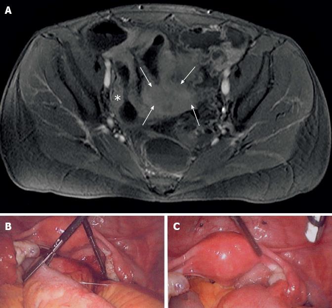
Figure 1 Radiological and surgical aspects of florid intestinal endometriosis and Crohn’s disease coincidence.
A: Magnetic resonance imaging-Sellink of the lower pelvis (axial T1 FS plus contrast agent). Bauhin’s valve and coecum are marked with a asterisk. The arrows highlight an uncharacteristic tissue mass involving the bowel; B: Laparoscopically, adhesion of the distal small bowel to the pelvis wall; C: Brownish spots on the pelvis and bladder peritoneum are indicating for endometriosis.
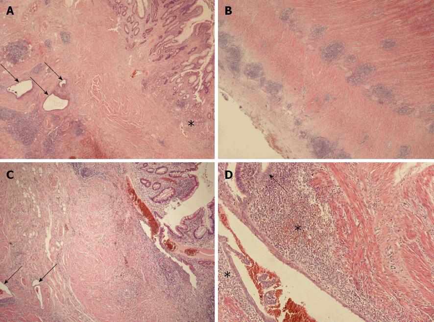
Figure 2 Coincidence of florid intestinal endometriosis and Crohn’s disease in small intestinal wall.
A: Complex inflammatory mucosal injury with establishment of pyloric gland metaplasia (asterisk). In deeper layers severe lymphoid aggregates (arrows) accompanied by endometrial glands exist (HE, × 10); B: Strong lymphoid hyperplasia partially affecting the prominent neuronal plexus, and serositis (HE, × 10); C: Florid aphthous mucosal lesion (HE, × 10), the arrows indicate endometriosis. D: Higher magnification of intestinal endometriosis(HE, × 100). Examples of epithelial proliferates are marked by an arrow. The cytogenic stroma is highlighted by asterisks. Note the intraglandular aggregates of erythrocytes.
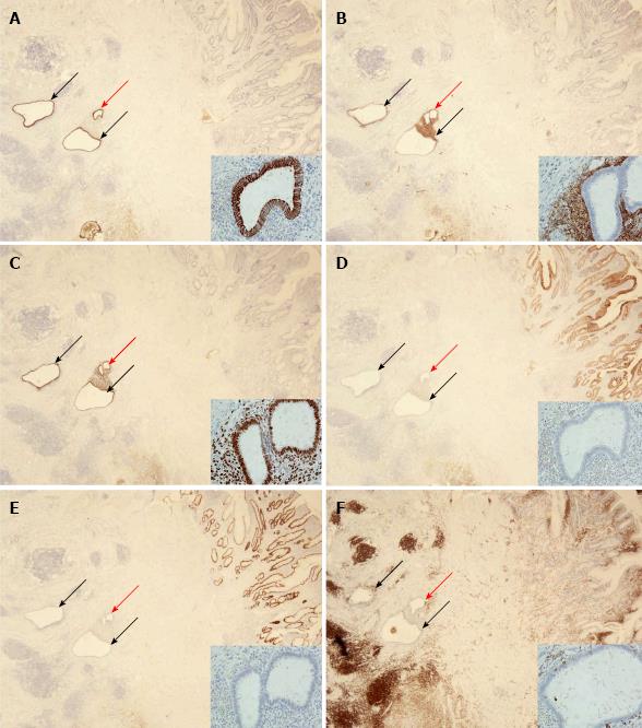
Figure 3 Immunohistochemical characterization of florid intestinal endometriosis and Crohn’s disease.
The intramural glandular structures (arrows in A-F) are positive for keratin 7 (A), CD10 (B), and progesterone receptor (C), but negative for keratin 20 (D) and CDX2 (E) which is vice versa to the mucosal staining; Strong anti-CD45 immunostaining illustrates severe lymphoid hyperplasia in CD (F). A-F: To illustrate immunostainings in detail always the intramural gland designated by a red arrow is shown in insets (original magnification: × 10; inset: × 250).
- Citation: Kaemmerer E, Westerkamp M, Kasperk R, Niepmann G, Scherer A, Gassler N. Coincidence of active Crohn's disease and florid endometriosis in the terminal ileum: A case report. World J Gastroenterol 2013; 19(27): 4413-4417
- URL: https://www.wjgnet.com/1007-9327/full/v19/i27/4413.htm
- DOI: https://dx.doi.org/10.3748/wjg.v19.i27.4413


