Copyright
©2013 Baishideng Publishing Group Co.
World J Gastroenterol. Jul 14, 2013; 19(26): 4267-4270
Published online Jul 14, 2013. doi: 10.3748/wjg.v19.i26.4267
Published online Jul 14, 2013. doi: 10.3748/wjg.v19.i26.4267
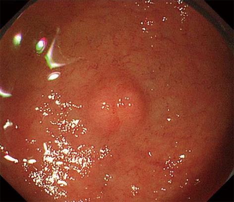
Figure 1 A neuroendocrine tumor, 5 mm in diameter, was detected in the duodenal bulb during endoscopy in an asymptomatic 57-year-old man.
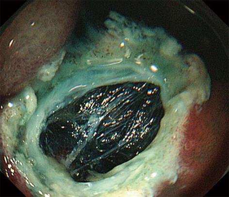
Figure 2 Although a blue-colored layer was identified in the resection defect, a small amount of a whitish layer was detected above the blue layer.
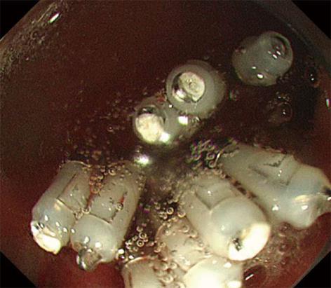
Figure 3 The defect was immediately closed with endoclips.
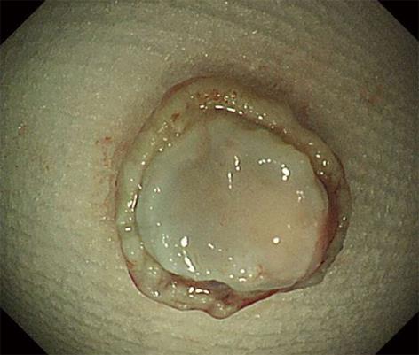
Figure 4 The muscle layer was clearly located on the underside of the resected specimen.
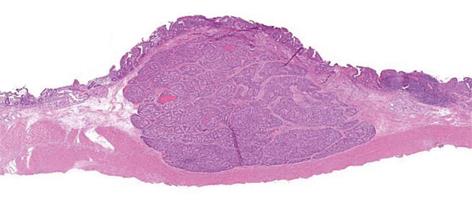
Figure 5 The muscularis propria was detected just beneath the submucosal layer (hematoxylin-eosin staining, loupe image).
- Citation: Hatogai K, Oono Y, Fu KI, Odagaki T, Ikematsu H, Kojima T, Yano T, Kaneko K. Unexpected endoscopic full-thickness resection of a duodenal neuroendocrine tumor. World J Gastroenterol 2013; 19(26): 4267-4270
- URL: https://www.wjgnet.com/1007-9327/full/v19/i26/4267.htm
- DOI: https://dx.doi.org/10.3748/wjg.v19.i26.4267


