Copyright
©2013 Baishideng Publishing Group Co.
World J Gastroenterol. Jul 14, 2013; 19(26): 4209-4213
Published online Jul 14, 2013. doi: 10.3748/wjg.v19.i26.4209
Published online Jul 14, 2013. doi: 10.3748/wjg.v19.i26.4209
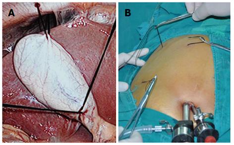
Figure 1 Suture suspension.
A: The fundus and Hartmann’s pouch were punctured and retracted by two sutures to expose Calot’s triangle; B: Puncture spot at the superior chest wall along the costal margin in order to draw the liver up a bit more.
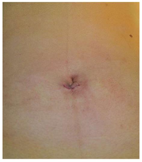
Figure 2 Umbilical incision was closed.
-
Citation: Cheng Y, Jiang ZS, Xu XP, Zhang Z, Xu TC, Zhou CJ, Qin JS, He GL, Gao Y, Pan MX. Laparoendoscopic single-site cholecystectomy
vs three-port laparoscopic cholecystectomy: A large-scale retrospective study. World J Gastroenterol 2013; 19(26): 4209-4213 - URL: https://www.wjgnet.com/1007-9327/full/v19/i26/4209.htm
- DOI: https://dx.doi.org/10.3748/wjg.v19.i26.4209


