Copyright
©2013 Baishideng Publishing Group Co.
World J Gastroenterol. Jul 7, 2013; 19(25): 4091-4093
Published online Jul 7, 2013. doi: 10.3748/wjg.v19.i25.4091
Published online Jul 7, 2013. doi: 10.3748/wjg.v19.i25.4091
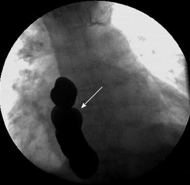
Figure 1 A roentgenogram showing foreign bodies in the lower esophagus (arrow).
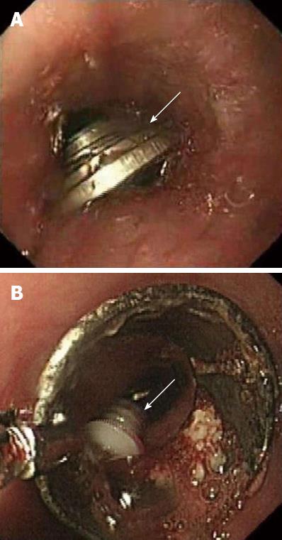
Figure 2 Gastroscope examination.
A: Gastroscopy displayed several coins (arrow) in the lower esophagus; B: A cylindrical plastic foreign body impacted in the esophageal wall (arrow).
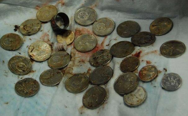
Figure 3 Foreign bodies after removal.
These included 26 coins, a ferrous ring, and a cylindrical plastic object.
- Citation: Li QP, Ge XX, Ji GZ, Fan ZN, Zhang FM, Wang Y, Miao L. Endoscopic retrieval of 28 foreign bodies in a 100-year-old female after attempted suicide. World J Gastroenterol 2013; 19(25): 4091-4093
- URL: https://www.wjgnet.com/1007-9327/full/v19/i25/4091.htm
- DOI: https://dx.doi.org/10.3748/wjg.v19.i25.4091


