Copyright
©2013 Baishideng Publishing Group Co.
World J Gastroenterol. Jul 7, 2013; 19(25): 4015-4022
Published online Jul 7, 2013. doi: 10.3748/wjg.v19.i25.4015
Published online Jul 7, 2013. doi: 10.3748/wjg.v19.i25.4015
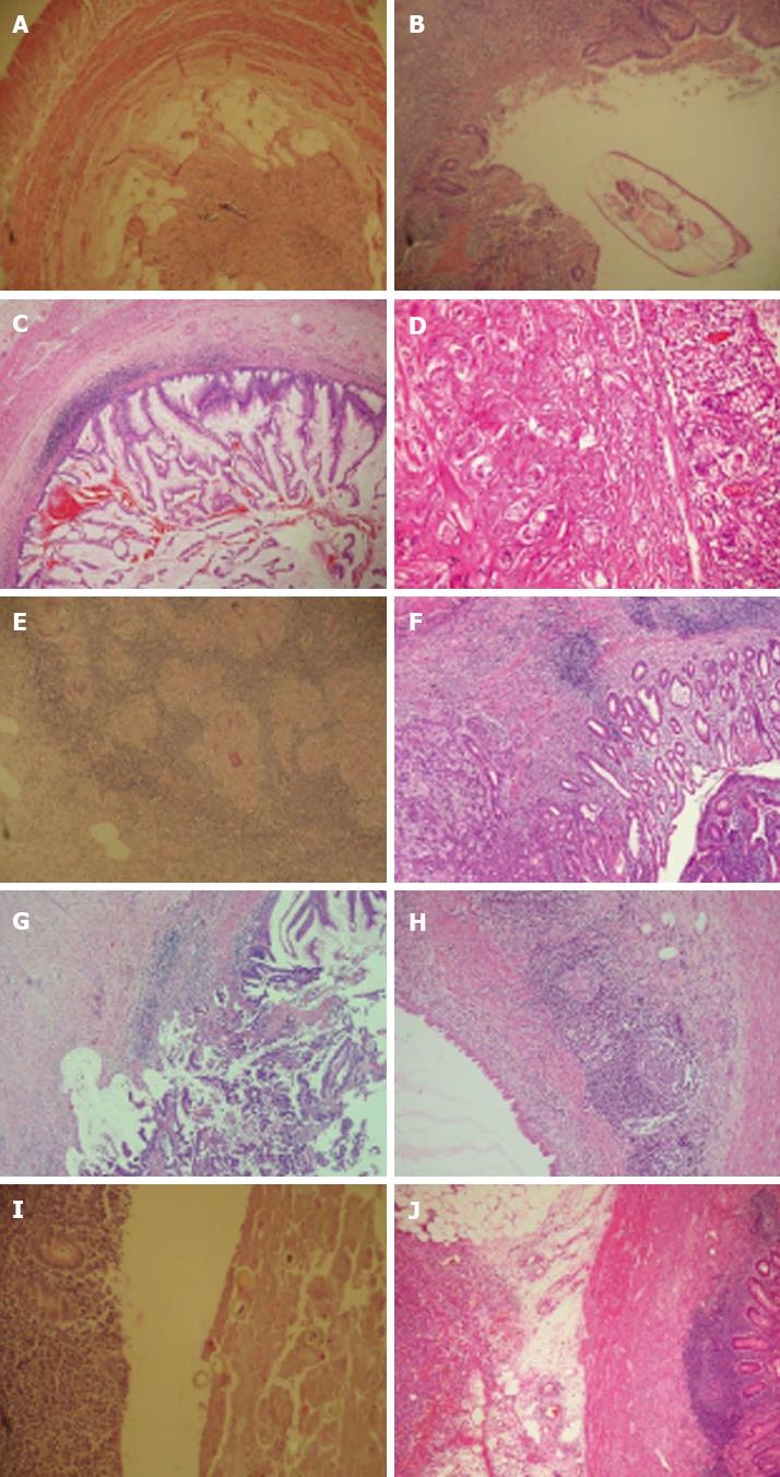
Figure 1 Unusual histopathologic findings.
A: Appendix vermiformis showing fibrous obliteration [hematoxylin and eosin (HE) × 40]; B: View of the enterobius vermiformis in the lumen of appendix vermiformis (HE × 100); C: Mucinous cystadenoma showing proliferation of neoplastic adenomatous epithelium, which exhibits low-grade dysplasia (HE × 100); D: Carcinoid tumor of the appendix showing rounded nests and tubules of tumor cells with uniform nuclei (HE × 200); E: Granulomatous inflammation. Submucosal granuloma with central necrosis (HE × 40); F: Moderately differentiated adenocarcinoma showing infiltration of the mucosa and submucosa of the appendiceal wall (HE × 100); G: Adenocarcinoma of the appendix showing associated mucocele on the top right side (HE × 100); H: Mucocele showing a unilocular dilated appendiceal wall lined with flattened epithelial cells (HE × 100); I: Eggs of Taenia sup are present in the lumen of appendix vermiformis (HE × 100); J: Serosa of the appendiceal wall showing diffuse large B cell lymphoma infiltration (HE × 40).
- Citation: Yilmaz M, Akbulut S, Kutluturk K, Sahin N, Arabaci E, Ara C, Yilmaz S. Unusual histopathological findings in appendectomy specimens from patients with suspected acute appendicitis. World J Gastroenterol 2013; 19(25): 4015-4022
- URL: https://www.wjgnet.com/1007-9327/full/v19/i25/4015.htm
- DOI: https://dx.doi.org/10.3748/wjg.v19.i25.4015


