Copyright
©2013 Baishideng Publishing Group Co.
World J Gastroenterol. Jul 7, 2013; 19(25): 3918-3930
Published online Jul 7, 2013. doi: 10.3748/wjg.v19.i25.3918
Published online Jul 7, 2013. doi: 10.3748/wjg.v19.i25.3918
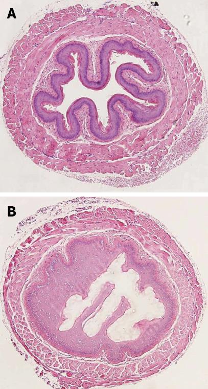
Figure 1 Murine esophagus exposed for 10 min to control (A) and 10% NaOH (B).
Reproduced from Osman et al[10].
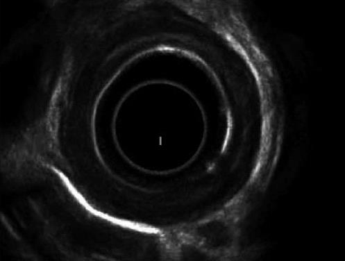
Figure 2 Endoscopic ultrasound showing involvement of the muscularis propria of esophageal wall.
Reproduced from Kamijo et al[37].
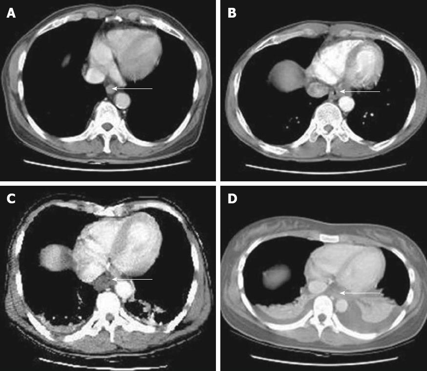
Figure 3 Computed tomography grading of esophageal caustic injuries.
A: Grade 1; B: Grade 2; C: Grade 3; D: Grade 4. Reproduced from Ryu et al[40]. Arrows show the esophageal wall.
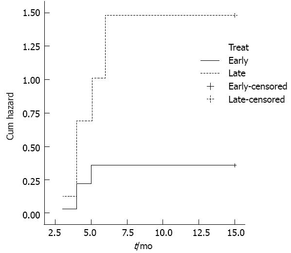
Figure 4 Significantly higher hazard of re-dilatation in patients submitted to late dilatation.
P = 0.0008. Reproduced from Contini et al[97].
- Citation: Contini S, Scarpignato C. Caustic injury of the upper gastrointestinal tract: A comprehensive review. World J Gastroenterol 2013; 19(25): 3918-3930
- URL: https://www.wjgnet.com/1007-9327/full/v19/i25/3918.htm
- DOI: https://dx.doi.org/10.3748/wjg.v19.i25.3918


