Copyright
©2013 Baishideng Publishing Group Co.
World J Gastroenterol. Jun 7, 2013; 19(21): 3364-3368
Published online Jun 7, 2013. doi: 10.3748/wjg.v19.i21.3364
Published online Jun 7, 2013. doi: 10.3748/wjg.v19.i21.3364
Figure 1 Computed tomography images of the abdomino-thoracic region showing a cystic mass in the liver with filiform calcification inside and a small amount of pleural effusion.
A: Plain; B: Contrast-enhanced; C: Reconstructive. RA: Right anterior.
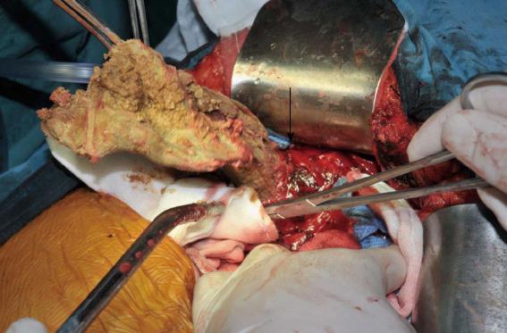
Figure 2 Surgical gauze (arrow) and the cyst wall (held with vessel forceps) in the right liver lobe following the evacuation of pus.
- Citation: Xu J, Wang H, Song ZW, Shen MD, Shi SH, Zhang W, Zhang M, Zheng SS. Foreign body retained in liver long after gauze packing. World J Gastroenterol 2013; 19(21): 3364-3368
- URL: https://www.wjgnet.com/1007-9327/full/v19/i21/3364.htm
- DOI: https://dx.doi.org/10.3748/wjg.v19.i21.3364


